Calcific tendinitis of the supraspinatus tendon
treated with iontophoresis: a case report
Sarvenaz Karimi-GhasemAbad 1 *, Alireza Rahmanizad 2
1 Razi Hospital, School of Medicine, Guilan University Medical
Sciences, Rasht, Iran
2 Physiotherapy Department of University of Social Welfare and
Rehabilitation Sciences, Tehran, Iran
Corresponding
Authors: Sarvenaz Karimi-GhasemAbad
* Email: s_karimi@gums.ac.ir
Abstract
Introduction: In particular, calcium hydroxyapatite crystals that are frequently
deposited within the supraspinatus and infraspinatus tendons are the cause of
calcific tendinitis of the shoulder, an acute or chronic painful condition
brought on by calcific deposits inside or around the rotator cuff tendons.
Case presentation: A 46-year-old patient arrived at the clinic complaining of
excruciating pain and significant movement impairment. Calcific tendonitis was
diagnosed during a clinical assessment. Iontophoresis using a 5% acetic acid
solution was applied three times a week for ten sessions as part of the
treatment.
Discussion: Various studies have identified 5% acetic acid iontophoresis as an
effective intervention for calcific tendinitis-associated pain. Additionally,
this treatment modality was partially responsible for the reduction in calcific
deposits.
Conclusion: Ten sessions of iontophoresis therapy using a 5% acetic acid solution
were conducted. Following completion, there was a full recovery of shoulder
range of motion, a complete clearance of calcific deposits, and no pain.
Keywords: Calcific tendinitis, Iontophoresis, Acetic acid, Shoulder
Key findings
1.
The pre-intervention
VAS for pain was 10/10; it decreased to 0/10 after the iontophoresis therapy
using a 5% acetic acid solution.
2.
The pre-treatment
range of motion was limited in all directions; the full range was regained
post-intervention.
3.
Complete resolution
of the calcific deposit that had been visible before treatment occurred after
iontophoresis therapy using a 5% acetic acid solution.
Introduction
The
self-limiting condition known as calcific tendinitis of the shoulder, or
enthesopathy, is typified by the accumulation of calcium phosphate crystals in
the rotator cuff tendons (1). It is uncommon in people over 70
and most frequently happens in those between the ages of 30 and 50 (2). It affects both shoulders in 10%
of patients, is more prevalent in the right shoulder than the left, and is
roughly twice as likely to occur in women as in males (3). The most frequent location is
1.5–2 cm from the greater tuberosity's supraspinatus tendon insertion point. It
has been reported in the literature that calcific tendinitis is more common in
the supraspinatus tendon than in other tendons (4).
The
way calcific tendinitis is treated varies, and a patient's level of pain is a
key consideration. The conventional treatment for calcific tendinitis often
starts with conservative management. If initial conservative measures prove
ineffective or under specific circumstances, surgical intervention may be
considered (5, 6). Recently,
various noninvasive and conservative treatment options have demonstrated
effectiveness. These include oral anti-inflammatory drugs, therapeutic
exercises, a combination of ultrasound therapy with therapeutic exercises,
Iontophoresis with acetic acid, ultrasound therapy paired with mesotherapy, and
ultrasound-guided needling (UGN) and extracorporeal shock wave therapy (ESWT) (7-9). These methods
have shown potential in reducing calcium deposit size, relieving pain, and
improving shoulder functionality (10).
Acetic
acid iontophoresis was first performed for the treatment of calcifying
tendinitis of the shoulder back in 1955. The physiological mechanism behind
this treatment is based on a transdermal drug delivery method wherein most
ionizable substances are transdermally delivered through the hair follicle and
sweat gland channels because of the polar action of direct-galvanic-current
motives. The resulting current, therefore, induces the translational motion of
ionized molecules positioned under the electrode of identical polarity toward
the electrode of opposite charge. In this specific arrangement, acetic acid-an
inorganic anion-was provided under the cathode or the negatively charged
electrode and was subjected to migration toward the positively charged electrode-the
anode-by the action of galvanic current. Since calcifications consist mainly of
hydroxyapatite crystals that are water-insoluble but acid-soluble, there is a
reasonable expectation of the reduction of calcification with this method (1). The effectiveness of 10 sessions
of 5% acetic acid iontophoresis in treating calcific tendinitis with a clinical
diagnosis is examined in this case report.
Case
presentation
A 46-year-old female working in the accounts section
in a children's hospital was referred for complaints of pain and stiff right
shoulder to the physiotherapy clinic. The patient reported that the shoulder
pain had gradually begun two months ago, leading to progressive limitation of
all shoulder movements. Activities of daily living, such as dressing and
eating, were notably affected.
Initial postural assessment during the course of the
examination revealed kyphotic posture with rounded shoulders and forward head
posture. On palpation, tenderness and pain on the shoulder were located over
the supraspinatus tendon, the deltoid, and subscapularis muscles. It was
impossible to perform a complete physical examination of the patient, including
active and passive range of motion assessments (The range of motion for
abduction and flexion was recorded at 20 degrees, with no observable extension or
rotational movement.) and specialized tests, in view of the patient's severe
pain, which he described as VAS scores of 10/10. Imaging finding [X-ray]
revealed calcification within the subacromial space (Figure 1).
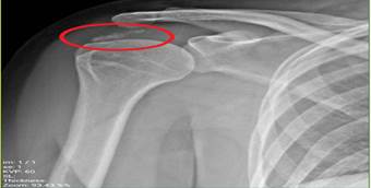
Figure
1.
Anteroposterior external rotation radiograph of the right shoulder,
demonstrating marked soft tissue calcifications identified at the insertion
site of the supraspinatus tendon.
So, following the diagnosis, the selected modality
of treatment was acetic acid iontophoresis. Acetic acid, being negatively
charged, was transferred into the body using the cathode. Iontophoresis
treatment was administered in sessions of 10 series, three times a week on
alternate days for 15 minutes each.
The cathode was then soaked in a solution made of
0.05% acetic acid and placed over the proximal attachment of the supraspinatus
tendon (This region, situated near the insertion of the superior rotator cuff,
is characterized by reduced vascularity(2)). It was to be wrapped in place with an
elastic bandage. The anode electrode was wet with water only and positioned on
the distal portion of the same hand over the bony region on the dorsal side of
the wrist. A galvanic current was delivered to using an Electrotherapy
stimulator.
The size of the cathode and anode pads used for the
treatment was each 20 cm². The position, according to Modalities for
Therapeutic Intervention (11) is that the maximum current density for the
cathode is 0.5 mA/cm² and for the anode is 1 mA/cm². With the size of the pad
being 20 cm², the maximum allowable current calculated to 10 mA. The current
ampitude was calculated using the following formulas (11):
![]()
The current amplitude in first two sessions was 6
mA, increasing in the third session to 8 mA and further increased in subsequent
sessions to 10 mA. During the first four sessions, manual release techniques
were also applied on the subscapularis and pectoralis minor muscles. By the end
of the fourth session, there was already a significant decrease in the
intensity of his pain, and he already had a VAS score of 2/10.
Starting with the fifth session, exercise teachings
related to kinesitherapy for kyphotic posture have been introduced in order to
further improve therapeutic results. At the end of the sessions, he had full
shoulder range in all movements and only felt pain at the end of the range of
motion. In the radiographic image, the calcific deposit from the supraspinatus
tendon insertion was gone (Figure 2).

Figure
2. Anteroposterior external rotation radiograph of the
right shoulder, demonstrates marked improvement in the previously noted soft
tissue calcifications identified at the insertion site of the supraspinatus
tendon.
Discussion
The
major treatment objectives for calcific tendinopathy include pain, limitation
of movement, and size of calcification (12). In the current study, all three
variables were measured as treatment outcomes. All three parameters showed
dramatic changes after iontophoresis with 5% acetic acid. In particular, the
patient's pain score reached zero by the end of ten sessions, the shoulder
range of motion became fully restored, and the calcification resolved
completely.
Acetic
acid iontophoresis has emerged as a promising conservative treatment for
calcifying tendinitis, particularly in the context of various tendons,
including the Achilles and shoulder (1). This technique utilizes a direct
electric current to enhance the transdermal delivery of acetic acid, which is
believed to facilitate the resorption of calcific deposits (13). While acetic acid iontophoresis
shows promise as an effective treatment for calcifying tendinitis, some studies
indicate that its efficacy may vary based on individual patient factors and the
specific tendon affected (1, 4, 14).
Iontophoresis
with 5% acetic acid has been shown in numerous studies to be effective in
lowering calcific tendinitis pain, it has been demonstrated that iontophoresis
with acetic acid not only lessens pain but also shrinks calcific deposits (15). In addition, in a case report, 3%
acetic acid was tested for its effect in calcific tendinopathy of the shoulder
for a period of 16 weeks. The results presented an agreement with the present
study; that also showed inadequate explanation for the effectiveness of
iontophoresis (12).
Although
interim analyses were promising, one cannot exclude that acetic acid does not
effectively penetrate into the skin barrier. Previous investigations carried
out on iontophoretic drug delivery did show the transdermal penetration of some
anti-inflammatory drugs and a poor passage of cortisone in humans; however, no
confirmation of transdermal absorption has been evidenced so far referring to
acetic acid itself(16, 17). Animal and human studies using
radioactive tracers, along with techniques such as fluorescein dye tracking and
scanning electrochemical microscopy, have explained the electrophoretic
processes: diffusion, migration, and electro-osmosis(18). None of these, however, have
actually researched resorptive capabilities of acetic acid through the skin
influenced by galvanic current(1). The lack of long-term follow-up
and the absence of comparisons with other therapeutic methods were among the
limitations of the present study.
Conclusion
While
galvanic stimulation chemical burns can be considered as some of the important
complications of iontophoresis, no such complication was observed in this case.
From the literature today, Extracorporeal shockwave therapy, Ultrasound-guided
needle lavage, and surgical intervention are the mainstream treatment for
shoulder calcium deposits (2). Only two clinical trials, so far,
have involved the use of 5% acetic acid iontophoresis for this condition. Both
used the primary outcomes as pain reduction and improvement in shoulder
function. By contrast, the therapeutic approach presented in the current study
brought about complete pain relief, full recovery of the range of motion of the
shoulder, normal function, and even complete disappearance of calcium deposits
that were confirmed with imaging after only ten sessions of treatment. Future
study could involve investigating the effects of acetic acid on skin
permeability and conducting clinical trial studies. And also, the comparison of
iontophoresis, shockwave therapy, and dry needling treatments could be explored
in future studies.
Author
contribution
SK-GA performed Conceptualization, Software, Methodology, Writing – Original,
performed Formal analysis. AR data collection.
Conflict
of interest
There
is no Conflicts of interest/competing interests.
Funding
There
is no funding.
References
1. Leduc BE, et al.
Treatment of calcifying tendinitis of the shoulder by acetic acid
iontophoresis: a double-blind randomized controlled trial. Archives of physical
medicine and rehabilitation. 2003;84(10):1523-7.
2. Suzuki K, et al. Calcific tendinitis of the
rotator cuff: management options. J Am Acad Orthop Surg. 2014;22(11):707-17.
3. Kim MS, et al. Diagnosis and treatment of
calcific tendinitis of the shoulder. Clin Shoulder Elb. 2020;23(4):210-6.
4. Aldehaim AY, Alarfaj AS. Global
Hypertrophic Calcification of Shoulder Joint Capsule. Clinical Medicine
Insights: Case Reports. 2021;14:11795476211025351.
5. Oh DG, Yoo KT. The effects of therapeutic
exercise using PNF on the size of calcium deposits, pain self-awareness, and
shoulder joint function in a calcific tendinitis patient: a case study. J Phys
Ther Sci. 2017;29(1):163-7.
6. Franceschi F, et al. Arthroscopic
management of calcific tendinitis of the subscapularis tendon. Knee Surgery,
Sports Traumatology, Arthroscopy. 2007;15(12):1482-5.
7. Scibek JS, Carcia CR. Presentation and
conservative management of acute calcific tendinopathy: a case study and
literature review. Journal of sport rehabilitation. 2012;21(4):334-42.
8. Abate M, et al. Usefulness of
rehabilitation in patients with rotator cuff calcific tendinopathy after
ultrasound-guided percutaneous treatment. Medical Principles and Practice.
2015;24(1):23-9.
9. Merolla G, et al. Calcific tendinitis of
the rotator cuff: state of the art in diagnosis and treatment. J Orthop
Traumatol. 2016;17(1):7-14.
10. Simpson M, et al. Effectiveness of
non-surgical interventions for rotator cuff calcific tendinopathy: A systematic
literature review. 2020.
11. Bellew JW, et al. Michlovitz's modalities for
therapeutic intervention: FA Davis; 2016.
12. Medina-Gandionco M, Briggs RA. Calcific
Tendinopathy of the Rotator Cuff Treated With Acetic Acid Iontophoresis. J
Orthop Sports Phys Ther. 2020;50(11):650.
13. Ciccone CD. Does acetic acid iontophoresis
accelerate the resorption of calcium deposits in calcific tendinitis of the
shoulder? Physical therapy. 2003;83(1):68-74.
14. Perron M, Malouin F. Acetic acid
lontophoresis and ultrasound for the treatment of calcifying tendinitis of the
shoulder: A randomized control trial. Archives of physical medicine and
rehabilitation. 1997;78(4):379-84.
15. Fernández-Cuadros ME, et al. Calcifying
tendonitis of the ankle, effectivenness of 5% acetic acid iontophoresis and
ultrasound over achiles tendon: A prospective case series. Int J Foot Ankle.
2019;3:023.
16. Chien YW, Banga AK. Iontophoretic
(transdermal) delivery of drugs: overview of historical development. Journal of
pharmaceutical sciences. 1989;78(5):353-4.
17. Chantraine Ae, et al. Is cortisone
iontophoresis possible? Archives of physical medicine and rehabilitation.
1986;67(1):38-40.
18. HT Z, et al. Iontophoresis studies with a
radioactive tracer. Archives of Physical Medicine and Rehabilitation.
1959;40(5):193-6.