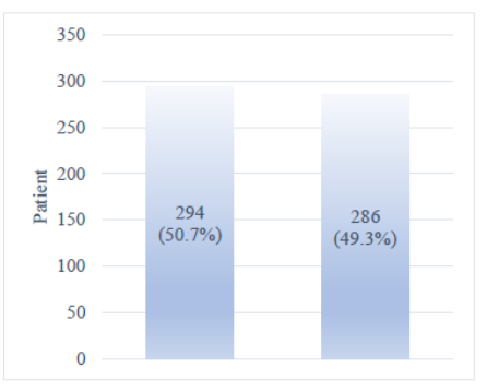
 Figure 1. Frequency distribution of gender of brain tumor patients examined in the study.
Introduction: Understanding the frequency of malignant and non-malignant brain tumors is crucial for understanding disease causes. Factors such as histological type, age of diagnosis, sex, and race are considered. Identifying risk factors such as allergy, ionizing radiation, and hereditary factors is important for prevention and early detection. Large epidemiological studies can provide a deeper understanding of this subject. Limited studies have been reported on the epidemiologic profile of brain tumors.
Materials and methods: This research was conducted on 580 patients, and all information on patients with malignant and benign brain tumors was extracted from their pathology reports, with emphasis on basic patient characteristics, such as age, gender, etc. All obtained data were statistically analyzed using software such as Excel and SPSS version 14, and the results were presented in the form of figures and tables.
Results: Gender distribution varied among different brain tumor groups and was reported as statistically significant. The frequency of malignant, benign, and uncertain or unknown behavior neoplasms was also studied by age. A significant relationship was found between age and the type of brain tumor. The frequency of different types of neoplasms according to the status of the patients showed significant differences between different brain tumor groups.
Conclusions: The highest frequency was attributed to unspecified or unknown neoplasms of the brain, followed by malignant neoplasms and benign neoplasms of the brain. The study found a statistically significant difference in age and sex among different tumor groups. Tumors were more common in women than in men, contrary to previous studies. The prevalence of surgical tumors in Rasht shows an increase.
Keywords: Brain tumors, Clinical and laboratory information, Frequency, Histopathology
It is crucial to look at all forms of brain tumors using data on incidence rates by demographic, including risk, gender, age, ethnicity, and other variables. We go over the many kinds of brain malignancies and tumors, as well as the available treatments today (1). Brain tumors are a constant source of concern for the global medical community, with over 300,000 instances recorded each year. Brain cancer can arise from brain tumors that have invaded the normal brain, even though some brain tumors may be benign. This page gives a summary of current research on brain tumors. It makes scientific facts readable for readers with varying backgrounds who are interested in learning more about this topic (2, 3).
Comprehending brain tumors is essential for improving public health since prevention begins with each person being aware of the dangers and symptoms of this condition. The general public can gain from learning about the findings of brain tumor research at the same time that medical professionals and neurosurgeons strive to enhance treatment options and prognoses for patients with these conditions. The use of artificial intelligence (AI) in medicine and other rapid technical breakthroughs has made it possible for clinicians to diagnose and predict the occurrence of brain tumors with ever-greater confidence and speed.
The vast majority of primary tumors that develop within the central nervous system (CNS) are gliomas. A "glioma" is a term used to describe tumors that resemble normal glial cells in histology, such as astrocytes, oligodendrocytes, and ependymal cells. These glioma categories all contain neoplasms with varying degrees of biological aggressiveness. In the past, World Health Organization (WHO) grades 1 and 2, which are slower-growing lesions, have been referred to as "low-grade gliomas," while grade 3 and 4 tumors, which progress more quickly, are referred to as "high-grade gliomas." Since these names combine several types of cancers, many of which have drastically different biological characteristics, prognoses, and treatment modalities, the WHO classification advises against using them (4-6).
For instance, the current classification of grade 1 and grade 2 gliomas emphasizes differentiating between more restricted astrocytic tumors (such as grade 1 pilocytic astrocytoma) and diffuse gliomas (such as oligodendroglioma and isocitrate dehydrogenase [IDH] mutant astrocytoma). Furthermore, the courses of grade 3 and grade 4 tumors can differ significantly, including how they respond to treatment. Neural progenitor cells are most likely the source of high-grade gliomas; however, it is unclear at what time these target cells differentiate into stem cells versus progenitor cells. Mouse models and molecular genetic analyses of tumor samples from patients, neighboring subventricular zone (SVZ) biopsies, and normal tissue in isocitrate dehydrogenase (IDH)-wildtype glioblastoma patients indicate that astrocyte-like neural stem cells in the SVZ, which have low-level somatic driver mutations, are the cell of origin in this disease (6).
These cells have the potential to move and pick up other somatic mutations over time, which could eventually result in the development of IDH-wildtype glioblastoma in other parts of the brain. Multipotent tumor stem cells found in high-grade gliomas are in charge of proliferating and repopulating the tumors (7-10). Since therapies that do not ablate the tumor stem cells will not be successful in eliminating the tumor, the existence of these tumor stem cells may have therapeutic consequences.
It is believed that particular genes on a cell's chromosomes become damaged and cease to function correctly, leading to brain tumors. These genes often control the pace of cell division, if any, and they also correct genes that cause other genes to malfunction, as well as those that should lead to the cell's self-destruction if damage is too great to be repaired. A person may occasionally be partially defective in one or more of these genes from birth. Subsequent environmental conditions could cause additional harm. In other situations, the only possible explanation could be environmental damage to the genes. Why some individuals in a given "environment" have brain tumors while others do not is unknown. A cell can eventually develop into a tumor if it is growing at a high rate and internal controls that regulate its growth are compromised (11-13).
The immune system of the body may provide an additional line of protection; in an ideal world, it would recognize the aberrant cell and eliminate it. Tumors have the ability to create chemicals that prevent the immune system from identifying the abnormal tumor cells, ultimately overwhelming all external and internal barriers to the tumor's continued growth. A fast-growing tumor could require more nutrients and oxygen than the nearby blood flow meant for healthy tissue can offer. Angiogenesis factors are chemicals that tumors can make to help blood vessels expand. The tumor receives more nutrition from the growing new vessels, and eventually, the tumor becomes reliant on them (14). Although some studies are being done in this area, further investigation is required to convert the findings into possible treatments.
Benign brain tumor kinds are categorized as follows. Most common in individuals between the ages of 50 and 60, chordomas are benign, slowly developing tumors. The base of the skull and the bottom part of the spine are where they are most frequently found. Rare tumors, accounting for only 0.2 percent of all primary brain tumors, craniopharyngiomas are usually benign but difficult to remove due to their location near important structures deep within the brain. Typically, they arise from a portion of the pituitary gland (the structure that regulates many hormones in the body), so nearly all patients will require some form of hormone replacement therapy (15, 16). Despite being benign, these tumors may invade the adjacent bone and put pressure on nearby neural tissue.
Rare tumors known as gangliocytomas, gangliomas, and anaplastic gangliogliomas, which mostly affect young adults, comprise neoplastic nerve cells that are rather well-differentiated. Usually benign, Globus jugular tumors are found at the top of the jugular vein, directly beneath the base of the skull. These are the most prevalent types of glomus tumors. But generally speaking, glomus tumors account for only 0.6% of head and neck neoplasms (17). Meningiomas represent 10 to 15 percent of all brain neoplasms, with a very tiny fraction becoming malignant. Meningiomas are the most prevalent benign intracranial tumors. The meninges, which are membrane-like structures that encircle the brain and spinal cord, are the source of these malignancies.
Pineocytomas are primarily adult-onset benign tumors that originate from the pineal cells. Most of the time, they are homogenous, slowly developing, well-defined, and noninvasive. Adenomas are by far the most common disease affecting the pituitary; they commonly affect people in their 30s or 40s, although they are also diagnosed in children (18). Most of these tumors can be successfully treated. Schwannomas are common benign brain tumors in adults. They arise along nerves, comprised of cells that normally provide the “electrical insulation” for the nerve cells. Pituitary adenomas are the most common intracranial tumors after gliomas, meningiomas, and schwannomas. The vast majority of pituitary adenomas are benign and fairly slow-growing. Even malignant pituitary tumors rarely spread to other parts of the body (19). Rather than invade the remaining normal nerve, schwannomas frequently cause it to move.
The most prevalent type of schwannoma is acoustic neuromas, which originate from the vestibulocochlear nerve, the eighth cranial nerve, which connects the brain to the auditory organs. These tumors are benign, but if they expand and put pressure on nerves and eventually the brain, they can result in life-threatening complications or even death. Other places include along nerves that supply the limbs and, less frequently, along the spine.
Crucially, malignant brain tumor kinds are categorized as follows:
In the meanwhile, Different Kinds of Brain Tumors are Tumors with a moderate growth rate, hemangioblastomas are typically seen in the cerebellum. They can be big, come from blood vessels, and frequently have a cyst attached to them. Men are more likely than women to have these tumors, which are more common in those between the ages of 40 and 60. Rare and extremely aggressive, rhabdoid tumors often spread throughout the central nervous system (20-22). They frequently show up in many bodily parts, particularly the kidneys. Though they can happen to adults as well, they are more common in young children. In Poursina Hospital in Rasht, Iran, we examined the frequency of all tumor kinds based on pathological criteria for this study.
This study is an analytical cross-sectional study conducted on patients with brain tumors admitted to Porsina Hospital in Rasht City, Gilan province, from 1399 to 1402. The study involved accessing the pathology report of patient samples from the pathology department of Porsina Hospital. Information from the files was collected based on the study checklist and entered into SPSS software version 25 for statistical analysis.
The data was analyzed using SPSS version 22 software. We used the Chi-square and Pearson tests to compare qualitative variables between different groups. For comparing quantitative variables between different groups, we used the ANOVA test and the T-test. In cases where normal distribution was lacking, we utilized the Kruskal-Wallis test.
The gender-based investigation in the study revealed the frequency distribution of patients with brain tumors. Out of 580 patients with brain tumors examined, 286 (49.3%) were male, and 294 (50.7%) were female (Figure 1).
Figure 1. Frequency distribution of gender of brain tumor patients examined in the study.
The frequency distribution of patients with brain tumors was investigated in the study based on age. Out of the 580 patients with brain tumors studied, the distribution by age is as follows: 10 (1.72%) were less than 10 years old, 8 (1.4%) were between 10 and 19 years old, 32 (5.51%) were between 20 and 29 years old, 79 (13.62%) were between 30 and 39 years old, 104 (17.93%) were between 40 and 49 years old, 135 (23.3%) were between 50 and 59 years old, 135 (23.3%) were between 60 and 69 years old, 56 (9.6%) were between 70 and 79 years old, and 21 (3.62%) were between 80 and 89 years old. The average age was 54 ± 16.35 years, and the youngest patient was 1 year old, while the oldest was 89 years old (Figure 2).
 Figure 2. Age frequency distribution of brain tumor patients examined in the study.
Out of 580 patients with brain tumors examined, there were 126 (21.72%) women and 143 (24.66%) men with malignant neoplasms, 57 (9.83%) women and 29 (5%) men with neoplasms categorized as benign, and 108 (18.62%) women and 117 (20.17%) men with unspecified or unknown neoplasms. The gender distribution varied significantly among the different brain tumor groups, as reported at a statistically significant level (P-value=0.012) (Table 1, 2 and 3) (Figure 3-14).
| Pathology Group | Specific Site / Type | Female | Male | Total | P-value |
|---|---|---|---|---|---|
| Malignant neoplasm | Cerebral meninges | - | 1 | 1 | 0.012 |
| Meninges, indeterminate | 7 | 1 | 8 | ||
| Malignant brain neoplasm | 1 | - | 1 | ||
| The brain, except for the lobes and ventricles | - | 2 | 2 | ||
| Frontal lobe | 5 | 4 | 9 | ||
| Temporal lobe | 5 | 6 | 11 | ||
| Partial lobe | - | 1 | 1 | ||
| Occipital lobe | 1 | 1 | 2 | ||
| Cerebral ventricle | - | 2 | 2 | ||
| Cerebellum | 7 | 7 | 14 | ||
| Brain stem | - | 4 | 4 | ||
| Brain overlap lesion | - | 1 | 1 | ||
| Brain, unspecified | 55 | 85 | 140 | ||
| Pituitary gland | - | 1 | 1 | ||
| Secondary malignant neoplasm of the cerebral and cerebral meninges | 47 | 27 | 74 | ||
| Benign neoplasm | Cerebral meninges | 18 | 4 | 22 | |
| Meninges, indeterminate | 5 | 2 | 7 | ||
| Cerebral, infratentorial | 8 | 5 | 13 | ||
| Brain, unspecified | 14 | 12 | 26 | ||
| Pituitary gland | 10 | 5 | 15 | ||
| Craniopharyngioma duct | 1 | - | 1 | ||
| Pineal gland | 1 | 1 | 2 | ||
| Neoplasm of uncertain or unknown behavior | The brain and central nervous system | 2 | 1 | 3 | |
| Cerebral, supratentorial | 1 | - | 1 | ||
| Cerebral, infratentorial | 2 | 1 | 3 | ||
| Brain, unspecified | 98 | 111 | 209 | ||
| Pituitary gland | 3 | 3 | 6 | ||
| Pineal gland | 2 | 1 | 3 |
 Figure 3. Explanation of the frequency of brain tumors according to gender in Porsina Hospital, Rasht, 2012-2018.
 Figure 4. Description of malignant neoplasms by gender at Porsina Hospital, Rasht, 1399-1402.
 Figure 5. Explanation of the abundance of benign neoplasms according to gender in Porsina Hospital, Rasht, 1402-1399.
 Figure 6. Explanation of the abundance of neoplasms of unknown or unknown behavior according to gender in Porsina Hospital in Rasht, 1399-1402.
| Pathology Group | Type / Sub-site | <10 | 10-19 | 20-29 | 30-39 | 40-49 | 50-59 | 60-69 | 70-79 | 80-89 | P-value |
|---|---|---|---|---|---|---|---|---|---|---|---|
| Malignant Neoplasm | Meninges of the Brain | 0 | 0 | 0 | 0 | 0 | 1 | 0 | 0 | 0 | 0.015 |
| Meninges, Unspecified | 0 | 0 | 0 | 1 | 1 | 1 | 3 | 0 | 2 | ||
| Malignant Brain Neoplasm | 0 | 0 | 0 | 0 | 0 | 0 | 1 | 0 | 0 | ||
| Brain, except Lobes and Ventricles | 0 | 0 | 0 | 0 | 0 | 0 | 0 | 2 | 0 | ||
| Frontal Lobe | 0 | 0 | 1 | 0 | 1 | 2 | 2 | 3 | 0 | ||
| Temporal Lobe | 0 | 1 | 1 | 2 | 1 | 1 | 4 | 0 | 1 | ||
| Parietal Lobe | 0 | 0 | 1 | 0 | 0 | 0 | 0 | 0 | 0 | ||
| Occipital Lobe | 0 | 0 | 0 | 0 | 0 | 1 | 0 | 1 | 0 | ||
| Brain Ventricle | 0 | 0 | 2 | 0 | 0 | 0 | 0 | 0 | 0 | ||
| Cerebellum | 2 | 1 | 0 | 1 | 2 | 2 | 5 | 1 | 0 | ||
| Brainstem | 0 | 1 | 1 | 0 | 0 | 1 | 1 | 0 | 0 | ||
| Brain Overlapping Lesion | 0 | 0 | 0 | 0 | 0 | 0 | 0 | 1 | 0 | ||
| Brain, Unspecified | 3 | 1 | 12 | 21 | 24 | 39 | 27 | 11 | 2 | ||
| Secondary Malignant Neoplasm | 0 | 0 | 1 | 10 | 17 | 23 | 17 | 5 | 1 | ||
| Benign Neoplasm | Meninges of the Brain | 0 | 0 | 0 | 0 | 8 | 5 | 6 | 2 | 0 | |
| Meninges, Unspecified | 0 | 0 | 0 | 0 | 2 | 3 | 7 | 1 | 0 | ||
| Brain, Infratentorial | 0 | 0 | 1 | 2 | 2 | 2 | 5 | 1 | 0 | ||
| Brain, Unspecified | 0 | 0 | 3 | 0 | 8 | 6 | 3 | 5 | 1 | ||
| Pituitary Gland | 0 | 0 | 1 | 3 | 3 | 4 | 3 | 1 | 0 | ||
| Craniopharyngioma Duct | 0 | 0 | 0 | 0 | 1 | 0 | 0 | 0 | 0 | ||
| Pineal Gland | 0 | 0 | 0 | 0 | 1 | 1 | 0 | 0 | 0 | ||
| Neoplasm of Uncertain or Unknown Behavior | Brain and Central Nervous System | 0 | 0 | 0 | 0 | 0 | 1 | 1 | 0 | 1 | |
| Brain, Supratentorial | 0 | 0 | 0 | 0 | 0 | 0 | 0 | 0 | 1 | ||
| Brain, Infratentorial | 0 | 0 | 0 | 0 | 0 | 1 | 2 | 0 | 0 | ||
| Brain, Unspecified | 5 | 4 | 7 | 31 | 32 | 47 | 55 | 18 | 10 | ||
| Pituitary Gland | 0 | 0 | 0 | 1 | 1 | 1 | 1 | 1 | 1 | ||
| Pineal Gland | 0 | 0 | 1 | 2 | 0 | 0 | 0 | 0 | 0 |
 Figure 7. Explanation of the frequency of brain tumors according to age in Porsina Hospital, Rasht, 1399-1402.
 Figure 8. Explanation of the frequency of malignant neoplasm according to age in Porsina Hospital, Rasht, 1402-1399.
 Figure 9. Explanation of the frequency of benign neoplasms according to age in Porsina Hospital, Rasht, 1402-1399.
| Pathology Group | Type / Location | Recovery | Partial Recovery | Personal Satisfaction | Follow-up | Death | Transfer | P-value |
|---|---|---|---|---|---|---|---|---|
| Malignant Neoplasm | Meninges of the Brain | 0 | 1 | 0 | 0 | 0 | 0 | 0.027 |
| Meninges, Unspecified | 0 | 6 | 1 | 0 | 1 | 0 | ||
| Malignant Brain Neoplasm | 0 | 1 | 0 | 0 | 0 | 0 | ||
| Brain, except Lobes and Ventricles | 0 | 0 | 0 | 1 | 1 | 0 | ||
| Frontal Lobe | 0 | 3 | 0 | 4 | 2 | 0 | ||
| Temporal Lobe | 0 | 3 | 0 | 4 | 4 | 0 | ||
| Parietal Lobe | 0 | 0 | 0 | 1 | 0 | 0 | ||
| Occipital Lobe | 0 | 0 | 0 | 1 | 0 | 0 | ||
| Brain Ventricle | 0 | 0 | 0 | 1 | 1 | 0 | ||
| Cerebellum | 0 | 5 | 1 | 2 | 6 | 0 | ||
| Brainstem | 0 | 1 | 0 | 1 | 2 | 0 | ||
| Brain Overlapping Lesion | 0 | 0 | 0 | 0 | 1 | 0 | ||
| Brain, Unspecified | 5 | 52 | 7 | 32 | 44 | 0 | ||
| Pituitary Gland | 0 | 0 | 0 | 0 | 1 | 0 | ||
| Secondary Malignant Neoplasm of Brain and Meninges | 4 | 24 | 10 | 1 | 34 | 1 | ||
| Benign Neoplasm | Meninges of the Brain | 0 | 12 | 3 | 2 | 5 | 0 | |
| Meninges, Unspecified | 0 | 3 | 0 | 0 | 4 | 0 | ||
| Brain, Infratentorial | 0 | 6 | 1 | 3 | 3 | 0 | ||
| Brain, Unspecified | 0 | 12 | 3 | 5 | 5 | 0 | ||
| Pituitary Gland | 1 | 5 | 1 | 1 | 7 | 0 | ||
| Craniopharyngioma Duct | 0 | 0 | 0 | 1 | 0 | 0 | ||
| Pineal Gland | 0 | 0 | 0 | 1 | 1 | 0 | ||
| Neoplasm of Uncertain or Unknown Behavior | Brain and Central Nervous System | 0 | 1 | 1 | 0 | 1 | 0 | |
| Brain, Supratentorial | 0 | 1 | 0 | 0 | 0 | 0 | ||
| Brain, Infratentorial | 0 | 2 | 0 | 0 | 1 | 0 | ||
| Brain, Unspecified | 4 | 102 | 30 | 13 | 60 | 0 | ||
| Pituitary Gland | 0 | 0 | 2 | 0 | 4 | 0 | ||
| Pineal Gland | 0 | 2 | 1 | 0 | 0 | 0 |
 Figure 10. Explanation of the frequency of neoplasms of unspecified or unknown behavior according to age in Porsina Hospital in Rasht, 1402-1399.
 Figure 11. Explanation of the frequency of brain tumors according to status in Porsina Hospital, Rasht, 1399-1402.
 Figure 12. Explanation of the frequency of malignant neoplasm according to the condition in Porsina Hospital, Rasht, 1402-1399.
 Figure 13. Explanation of the frequency of benign neoplasms according to the condition in Porsina Hospital, Rasht, 1402-1399.
 Figure 14. Explanation of the frequency of unspecified or unknown neoplasms according to status in Porsina Hospital, Rasht, 1402-1399.
Out of the 580 patients with brain tumors that were studied, the patients were classified based on their age and the type of tumor (malignant, benign, unspecified, or unknown) as shown in the table below. The average age across the different groups was 54 years with a standard deviation of 16.35 years, and there was a significant difference in age distribution among the different brain tumor groups. (P-value=0.015)
Out of 580 patients with brain tumor investigated, in patients with malignant neoplasm, the condition of the patients included 9 (1.55%) patients with recovery, 96 (16.55%) patients with partial recovery, 19 (3.28%) patients With personal consent, 48 (28.8%) patients were being followed up, 97 (16.72%) patients died and 1 (0.17%) patient was transferred.
While in benign neoplasm patients, 1 (0.17%) patient with recovery, 38 (6.55%) patients with partial recovery, 8 (1.38%) patients with personal satisfaction, 13 (2.24%) The patient is being followed up and 25 (4.31%) patients have died, and also 4 (0.69%) patients with recovery, 108 (18.62%) patients with partial recovery, 34 (8.5%) patients with personal consent, 13 (2.24%) patients under follow-up, and 66 (11.38%) deceased patients were reported for unspecified or unknown neoplasm. There was a significant difference in the condition of patients between different brain tumor groups. (P-value=0.027).
This study aimed to investigate the frequency of brain tumors in Rasht's Porsina Hospital based on pathological characteristics in the years 2019 to 2019. 580 cases of brain tumors were diagnosed in Porsina Hospital, Rasht, in the Pathology Department. As mentioned in the findings section, the prevalence of brain tumors in affected people according to gender was 286 (49.3%) men and 294 (50.7%) women. In this regard, the results show that the number of infected people is slightly higher in women than in men. These findings do not agree with the statistics available in pathology sources. Because most sources have reported that brain tumors are more common in men than women, in this regard, another similar study was conducted by Albasri Abdulkader Mohammed and colleagues, who described the histopathological types and primary demographic parameters of brain tumors in Saudi Arabia and analyzed and compared the findings with previously published articles.
The result of their research was that a total of 227 brain tumors were registered in 122 (53.7%) men and 105 (46.3%) women patients. Pediatric and adult patients accounted for 10.6 and 89.4% of cases, respectively. The predominant age group of patients was between 40 and 49 years old (23.5%). The most common histopathological diagnosis in the present study was meningioma (30.8%), astrocytic tumors (29.1%), metastatic tumors (7.7%), and embryonal tumors (6.6%). Meningothelial meningioma was the most common type of meningioma (48.5%). The majority of astrocytic tumors (52%) were classified under WHO grade IV (19).
While in this study, according to the age of 580 patients with tumors, the lowest affected age is between 10-19 years old and the highest affected age is 50-59 and 60-69 according to graph 2 and Also, in this study, the frequency of the relationship between gender and the type of brain tumors was different and this difference was reported to be statistically significant. In patients with malignant neoplasm (brain, unspecified), the number of men is more than women, and the number of malignant neoplasm (cerebellum) is equal to that of men, and the meninges of the brain, partial lobe, occipital lobe, and pituitary gland are the least number among men and women in this study. And secondary malignant neoplasm (brain meninges) was more common in female patients in this study, which is consistent with reference books.
In this study, patients with benign neoplasms were more common in women according to gender, and also patients with neoplasms of unknown or unknown behavior were more common in men than women. The average age between different groups was 54 ± 16.35 years, and the age distribution between different brain tumor groups was significantly different, according to Table 3. In our study, patients aged 50-59 years were mostly undiagnosed with malignant neoplasms of the brain. Also, patients aged 40-49 had more benign neoplasms of the brain, unspecified, and benign neoplasms of the brain meninges. Patients aged 60-69 were more likely to have neoplasms of unknown or unknown brain behavior, unspecified.
In this regard, the relationship between the patient's condition and the type of brain tumor, in this study, the highest number was reported in malignant brain neoplasm, 5 patients recovered, 52 patients partially recovered, 7 patients were satisfied, 32 patients were followed up, and 44 patients died. Only 1 person was transferred with a benign neoplasm, and the largest number was in a benign neoplasm of the brain, unspecified, and meninges. 12 people got partial recovery, and only 1 person got better in a benign neoplasm of the pituitary gland, and the largest number in personal satisfaction. In benign neoplasm of the brain, unspecified and brain meninges, there were 3 people, and in benign neoplasm, the highest number of follow-ups related to the brain was unknown, which was 5 people, and the highest number of deaths in benign neoplasm was the pituitary gland, which was reported to be 7 people.
The highest number of recovery in neoplasm of unknown or unknown brain behavior was 4 people, and the highest number of relative recovery in neoplasm of unknown or unknown brain behavior was 102 people, and in this group, the highest personal satisfaction was 30 people, the highest follow-up statistics was 13 people, and the highest mortality rate was 60 was a person. And in total, the highest number of deaths was related to neoplasm of unknown or unknown behavior (brain, unknown) and the highest recovery was related to malignant brain neoplasm, unknown according to table 4.
In another similar study conducted by Abbas Rezaianzadeh, the results showed that 1043 cases of malignant brain neoplasm were recorded during the study period. Of these cases, 41.9% were women, and the incidence of the disease in men (15.26/100000) was higher than in women (19.45/100000). The incidence rate of malignant brain neoplasm showed an increasing trend from 2011 to 2013, but this trend was not statistically significant. This study did not investigate benign neoplasms and neoplasms of uncertain or unknown behavior (20).
In another study conducted by M. Mehrazin et al., like the current study, there was no significant relationship between sexes in terms of age, so Mehrazin et al. reported that meningioma was the most common histological type, followed by astrocytoma, pituitary adenoma. Glioblastoma and ependymoma were located. These account for 84% of all brain tumors. Male predominance was observed in several tumor types, while meningioma was more common in females. Brain tumors occurred in different age groups; 28% of cases occurred in patients less than 20 years old, 45% in patients 21 to 45 years old, 25% in patients 46 to 65 years old, and 2% in patients older than 65 years old. This study provided important epidemiological information about brain tumors in Iran (22).
Brain tumors are among the most common cases in the field of neuropathology. These tumors can be of different types, each with distinct pathological characteristics. Studying the frequency of these tumors based on their pathological characteristics can provide valuable insights into these diseases and their treatment methods. Previous research has indicated that the pathological characteristics of brain tumors can significantly impact the prognosis and treatment of these diseases. Therefore, this research has the potential to greatly contribute to progress in the field of neuropathology and improve the care of patients with brain tumors.
In Iran, there are many cases of brain neoplasms, but there have been few studies on the distribution of CNS tumors based on different histopathological characteristics. This study in Rasht highlights important pathological and epidemiological features of primary brain tumors, providing valuable preliminary information for future research. Given the impact of brain tumors on healthcare, social systems, and patients' psychological well-being, it is evident from this study that more aggressive types of brain malignancies are increasing. Therefore, it is necessary to conduct additional studies to identify potential risk factors and the role of current technology in addressing this disease. Given the significance of brain cancer frequency and its multifactorial nature, including environmental factors and pathological characteristics, early diagnosis, prevention, and effective treatment management are crucial. Therefore, the findings of this research can be extremely impactful.
AAS and PK conceived and designed the study and also wrote and edited the article. SARSh, KD, ZT, AA, accompanied many other parts of the manuscript, including data validity, writing, and scientific investigation. All authors read the manuscript comprehensively and confirmed the paper's final version.
There are no conflicts of interest.
We thank Guilan University of Medical Sciences for approving this project (IR.GUMS.REC.1402.463) and everyone who was involved in this project and contributed to it. In this way, we express our appreciation to Ms. Kosar Babaei for assisting us in coordinating some sections of this project.
There is no funding.