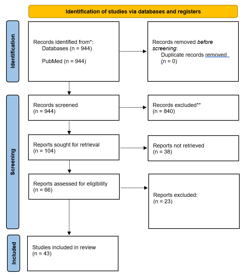
 Figure 1. PRISMA flow diagram.
Introduction: Immune checkpoint inhibitor-associated myocarditis (ICI-M) is a rare but life-threatening toxicity. This systematic review synthesizes the current evidence on the epidemiology, clinical presentation, diagnostic approaches, management strategies, and outcomes of ICI-M to guide clinical practice.
Materials and methods: We systematically searched PubMed from inception to January, 2026 for studies reporting on ICI-M in cancer patients. Data on patient demographics, clinical features, diagnostic findings, treatment, and outcomes were extracted. The risk of bias was assessed using appropriate tools.
Results: 43 studies were included. ICI-M predominantly affected older adults (median age 65-74 years) with metastatic melanoma, non-small cell lung cancer, or renal cell carcinoma. The highest risk was associated with combination ICI therapy (anti-PD-1/PD-L1 + anti-CTLA-4). Clinical presentation ranged from asymptomatic biomarker elevation to fulminant heart failure, with a high frequency of concurrent myositis. Key diagnostic findings included elevated troponin (>90% of cases), ECG abnormalities, and reduced global longitudinal strain on echocardiography. Management universally involved ICI discontinuation and high-dose corticosteroids. Second-line immunosuppression (e.g., IVIG, infliximab, abatacept) was used in refractory cases. Despite treatment, mortality remained high (25-50%). Poor prognostic factors included high troponin levels, reduced left ventricular ejection fraction, and conduction abnormalities.
Conclusion: ICI-M is a severe complication with high mortality. Early recognition via proactive monitoring, prompt diagnosis using a multi-modal approach, and immediate, aggressive immunosuppression are critical. Future research should focus on predictive biomarkers and randomized trials to optimize management.
Keywords: Immune checkpoint inhibitors, Myocarditis, Cardio-oncology, Immunotherapy, Immune-related adverse events, Systematic review
Immune checkpoint inhibitors (ICIs) have revolutionized oncology by harnessing the immune system to fight cancer. However, this enhanced immunity can also lead to immune-related adverse events (irAEs), affecting various organs. Among these, ICI-associated myocarditis (ICI-M) is one of the most severe, with a fatality rate exceeding many other irAEs (1, 2).
Despite its rarity, ICI-M presents a significant clinical challenge due to its non-specific presentation, rapid progression, and high mortality (3, 4). The clinical spectrum is broad, ranging from subclinical disease detected only by biomarker elevation to fulminant myocarditis and cardiogenic shock. Early diagnosis and intervention are paramount, yet standardized guidelines are still evolving based on accumulating evidence from cohort studies, registries, and case series.
Over the past decade, numerous studies have characterized the risk factors, clinical course, and outcomes of ICI-M. However, a comprehensive synthesis of this evidence is needed to consolidate our understanding and inform clinical decision-making. This systematic review aims to provide a detailed analysis of the global research landscape, patient characteristics, diagnostic findings, management strategies, and outcomes of ICI-M, integrating data from a wide range of published studies to offer a definitive overview for clinicians and researchers.
This systematic review was conducted and reported in accordance with the Preferred Reporting Items for Systematic Reviews and Meta-Analyses (PRISMA) guidelines.
A systematic search was performed in PubMed from database inception to January, 2026. The search strategy combined terms related to ("immune checkpoint inhibitor" OR "anti-PD-1" OR "anti-PD-L1" OR "anti-CTLA-4") AND ("myocarditis" OR "cardiotoxicity" OR "cardiovascular adverse event"). Proceedings from key cardiology and oncology conferences were also screened.
Studies were included if they: (1) reported on human cancer patients diagnosed with ICI-M; (2) provided original data on epidemiology, clinical presentation, diagnosis, management, or outcomes; and (3) were published in English. Case reports, cohort studies, registries, and clinical trials were eligible.
Two reviewers independently screened titles, abstracts, and full-text articles. Data were extracted using a standardized form, capturing information on study design, patient demographics, cancer types, ICI regimens, diagnostic criteria, management, and outcomes. The risk of bias for RCTs was assessed using the Cochrane Risk of Bias 2 (RoB 2) tool.
Given the heterogeneity in study designs and reporting, a narrative synthesis was conducted. Data are presented in summary tables and descriptive text.
The initial search yielded 944 records. After removing duplicates and screening titles and abstracts, 104 full-text articles were assessed for eligibility. Ultimately, 43 studies were included in the final synthesis (Figure 1).
Figure 1. PRISMA flow diagram.
The methodological quality of the included studies was assessed. The overall risk of bias was low to moderate. Common limitations included the retrospective nature of most studies and potential selection bias in single-center cohorts. The risk of bias summary and graph are presented in Figures 2a and 2b.
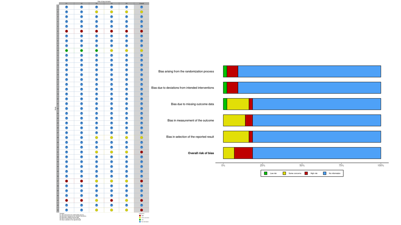 Figure 2. Risk of bias assessment across included studies. Figure 2a shows the proportion of studies assessed for various domains of bias, including: selection of participants, confounding variables, measurement of exposure, blinding of outcome assessment, incomplete outcome data, and selective outcome reporting. Each domain is color-coded to represent the assessed level of bias: Low risk (green), Unclear risk (yellow), High risk (red), Critical risk (dark red), and No information (blue). Figure 2b provides a study-wise breakdown of risk of bias assessments, allowing a granular comparison across individual studies.
The 43 included studies originated from a range of countries, with the United States (37.2%), China (20.9%), and Japan (14.0%) being the largest contributors (Table 1). This distribution highlights a significant geographical evidence gap, with vast regions like South America, Africa, and Eastern Europe unrepresented.
| Country | Number of Studies | Percentage of Total (n=43) | Study Types (Representative Examples) |
|---|---|---|---|
| United States (USA) | 16 | 37.2% | Multicenter registries, Single-center cohorts, Systematic Reviews, Case Reports, Preclinical/Translational |
| China | 9 | 20.9% | Retrospective multicenter cohorts, Single-center cohorts, Case Series, Bioinformatics analysis, Review Articles |
| Japan | 6 | 14.0% | Prospective observational studies, Retrospective cohort studies, Case Reports |
| France | 3 | 7.0% | Case-Control Studies, Case Reports with novel therapeutics |
| Germany | 2 | 4.7% | Prospective cohort, Preclinical/Clinical study |
| Multi-National | 2 | 4.7% | International retrospective cohort, International multicenter study |
| Australia | 1 | 2.3% | Case Report |
| Switzerland | 1 | 2.3% | Case Report |
| The Netherlands | 1 | 2.3% | Review Article |
| Canada | 1* | 2.3% | Part of a multicenter study |
| United Kingdom | 1* | 2.3% | Collaborator in a preclinical study |
| Austria | 1* | 2.3% | Collaborator in a preclinical study |
*Note: Studies where the country was a collaborator rather than the primary site.
This table provides a critical analysis of the global research landscape for ICI-associated myocarditis. The distribution is not uniform, revealing clear leaders and significant gaps. The dominance of the United States, contributing over a third of the studies, reflects its pioneering role in immuno-oncology, the high volume of patients treated at major cancer centers, and the early establishment of dedicated cardio-oncology research programs (3, 8, 11, 37, 39, 41). This leadership is evidenced by a diverse output, from large, foundational multicenter registries to cutting-edge translational science. China's position as the second-largest contributor signals its rapidly expanding capacity and focus in this field, often characterized by large-scale retrospective clinical cohorts that leverage its vast patient population to generate significant real-world evidence (12, 22, 23, 29, 30). Japan's significant output, while smaller in volume, is marked by high-quality, meticulous prospective and observational studies that have been instrumental in characterizing subclinical disease and detailed management (9, 16, 31, 35, 40, 43).
The European contributions, particularly from France and Germany, are notable for their highly specialized and innovative nature, such as pioneering the use of novel therapeutic agents like abatacept (5, 6) and producing key prospective biomarker studies (2). The presence of multi-national collaborations (2, 8) underscores the importance of pooling data to study this rare condition. Crucially, this map highlights a substantial evidence gap, with vast regions of the world (e.g., South America, Africa, Southeast Asia, Eastern Europe) unrepresented. This lack of geographical diversity may limit the generalizability of findings, as genetic backgrounds, regional cancer types, and healthcare delivery systems can influence the presentation and management of ICI-myocarditis.
The "typical" patient with ICI-M was an older adult (median age 65-74 years) with metastatic cancer, most commonly melanoma, non-small cell lung cancer (NSCLC), or renal cell carcinoma (RCC) (Table 2). Hypertension and diabetes were the most frequent comorbidities. A subset of patients had a pre-existing autoimmune condition.
This table synthesizes the demographic and clinical foundation of the studied population, painting a picture of the "typical" patient at risk for this severe toxicity. The consistency across numerous international cohorts strongly suggests that ICI-M, while rare, does not occur randomly but is more likely in a specific patient profile. The predominance of older patients (3, 11, 18, 37) likely reflects both the higher incidence of cancers treated with ICIs in this age group and a potentially altered immune response. The recurrence of melanoma, NSCLC, and RCC (2, 3, 8, 17) is expected, as these were among the first malignancies with approved ICI therapies, leading to extensive clinical experience and larger patient pools for observation. The overwhelming representation of metastatic disease (11, 16, 22, 30) is a critical confounder; it is unclear whether advanced cancer stage is an independent risk factor or if the association is simply because these patients are the primary recipients of ICIs. The high prevalence of hypertension and other cardiovascular comorbidities (3, 8, 12, 18) indicates that a compromised cardiovascular system may be more susceptible to immune-mediated injury, a key consideration for pre-therapy risk assessment.
| Characteristic | Summary Finding (Range or Most Frequent) | Details / Specifics | Key References |
|---|---|---|---|
| Median Age | 65 - 74 years | The patient population predominantly comprised older adults. | Mahmood et al., 2018 (65±13) (3); Dubey et al., 2025 (74 IQR 68-78) (11); Alexander et al., 2025 (74±9.4) (18); Puzanov et al., 2021 (73 IQR 66-79) (37) |
| Common Cancer Types | Melanoma, Lung Cancer, Renal Cell Carcinoma (RCC) | Melanoma was the most frequently reported cancer. | Lehmann et al., 2023 (Melanoma 20%, NSCLC 40%, RCC 10%) (2); Mahmood et al., 2018 (Melanoma 46%, NSCLC 11%) (3); Awadalla et al., 2020 (Melanoma 41%, Lung 16%, RCC 8%) (8); Jensen et al., 2025 (Melanoma, NSCLC, RCC) (17) |
| Cancer Stage | Predominantly Metastatic (Stage IV) | The vast majority of patients had advanced or metastatic disease. | Dubey et al., 2025 (Majority Stage IV) (11); Todo et al., 2025 (Metastatic) (16); Cao et al., 2025 (Stage III-IV NSCLC) (22); Tang et al., 2023 (71.6% Stage IV) (30) |
| Common Comorbidities | Hypertension, Diabetes Mellitus, Coronary Artery Disease | Hypertension was the most prevalent comorbidity. | Mahmood et al., 2018 (Hypertension 34%, Diabetes 34%) (3); Awadalla et al., 2020 (Hypertension 57%, Diabetes 24%) (8); Zheng et al., 2024 (Hypertension 38.6%, Diabetes 18.2%) (12); Alexander et al., 2025 (Hypertension 78%, Diabetes 28%) (18) |
| Pre-existing Autoimmune Condition | Present in a subset of patients | Noted in several case reports and cohorts. | Ganatra & Neilan, 2018 (Hashimoto's thyroiditis) (13); Stein-Merlob et al., 2021 (Graves' disease) (27) |
Combination ICI therapy (e.g., ipilimumab + nivolumab) was the strongest risk factor for ICI-M (Table 3). The median time to onset was early, typically within the first 1-4 cycles (17-65 days). A significant proportion of patients received concurrent therapies like chemotherapy or radiotherapy.
This table moves from who is at risk to what precipitates the event, highlighting modifiable risk factors. The strong, consistent signal across nearly all studies that combination ICI therapy (most notably anti-PD-1 + anti-CTLA-4) is the single greatest risk factor (1, 3, 7, 13, 17) is perhaps the most critical clinical finding. This makes biological sense, as dual checkpoint blockade induces a more potent and broader immune activation, inadvertently increasing the risk of breaking self-tolerance. The early median time to onset (3, 7, 16, 21, 32) underscores the need for heightened vigilance during the initial treatment cycles, fundamentally shaping monitoring guidelines. However, the reports of late-onset cases (21, 35, 41) remind clinicians that risk never completely dissipates. The data on concurrent therapies is more ambiguous, as many patients receive multi-modal treatment; while not definitively proven to be independent risk factors, chemotherapy and radiotherapy may create a pro-inflammatory environment that lowers the threshold for myocarditis (22, 30, 40).
| Factor | Summary Finding | Details / Specifics | Key References |
|---|---|---|---|
| Most Common ICI Classes | Anti-PD-1, Anti-PD-L1, Anti-CTLA-4 | Myocarditis was reported with all major ICI classes. | Moslehi et al., 2021 (1); Palaskas et al., 2020 (7); Turker & Johnson, 2023 (21) |
| Highest Risk Regimen | Combination ICI Therapy (e.g., Ipilimumab + Nivolumab) | Combination therapy was consistently identified as the strongest risk factor. | Moslehi et al., 2021 (1); Mahmood et al., 2018 (34% on combination) (3); Ganatra & Neilan, 2018 (13); Jensen et al., 2025 (17) |
| Median Time to Onset | 17 - 65 days | Onset was often early in the treatment course. | Mahmood et al., 2018 (34 days) (3); Palaskas et al., 2020 (27-65 days) (7); Todo et al., 2025 (25 days) (16); Turker & Johnson, 2023 (27-34 days) (21); Atallah-Yunes et al., 2019 (22.5 days) (32) |
| Typical Cycle of Onset | Within first 1-4 cycles | Many cases occurred after the first or second dose. | Atallah-Yunes et al., 2019 (1-2 doses in majority) (32); Cao et al., 2025 (48.4% within first 2 cycles) (22) |
| Concurrent Therapies | Chemotherapy, Targeted Therapy, Radiotherapy | A significant proportion received concurrent treatments. | Cao et al., 2025 (84.8% ICI combo with chemo) (22); Tang et al., 2023 (54.3% concurrent chemo) (30); Matsumoto et al., 2022 (prior radiotherapy) (40) |
The clinical presentation of ICI-M was highly variable (Table 4). Dyspnea was the most common symptom, but a significant proportion of patients were asymptomatic. Overlap with myositis and myasthenia gravis was frequent. Key diagnostic findings included elevated troponin, ECG abnormalities, and reduced global longitudinal strain (GLS) on echocardiography. Cardiac MRI and endomyocardial biopsy were crucial for confirmation. This table captures the clinical essence of the disease, revealing a spectrum from silent, biomarker-only disease to catastrophic failure. The high rate of asymptomatic or mildly symptomatic presentation (9, 16, 22, 31) is a pivotal finding; it argues strongly for proactive biomarker screening rather than relying on symptom reporting alone. The frequent overlap with myositis and myasthenia gravis (2, 5, 6, 23, 26) is a unique and dangerous feature, suggesting a shared antigenicity between cardiac, skeletal muscle, and neuromuscular junctions that is unmasked by ICIs. The diagnostic pillars are clearly established: Troponin is the cornerstone biomarker (3, 7, 8, 11), with its peak level being profoundly prognostic. Echocardiography often reveals preserved LVEF (3, 8, 26), but the superior sensitivity of GLS provides critical prognostic information even when LVEF appears normal (8, 9). Cardiac MRI is the best non-invasive tissue characterization tool (7, 13, 17), yet its false-negative rate confirms that it should complement, not replace, clinical judgment. The gold standard remains endomyocardial biopsy (3, 5, 13), which definitively reveals the T-cell-mediated pathology.
| Category | Finding | Details / Specifics | Key References |
|---|---|---|---|
| Common Symptoms | Dyspnea, Chest Pain, Fatigue | Dyspnea was the most common symptom. A significant proportion were asymptomatic. | Mahmood et al., 2018 (Dyspnea 71%) (3); Tanabe et al., 2021 (Asymptomatic) (9); Todo et al., 2025 (Some asymptomatic) (16); Cao et al., 2025 (45.5% symptomatic) (22); Nishikawa et al., 2022 (Mostly asymptomatic) (31) |
| Overlap Syndromes | Myositis, Myasthenia Gravis | Concomitant myositis was very common. | Lehmann et al., 2023 (~68% myositis) (2); Nguyen et al., 2022 (concurrent myositis) (5); Salem et al., 2019 (myositis) (6); Ke et al., 2023 (myositis & MG) (23); Sessums et al., 2020 (myositis) (26) |
| ECG Abnormalities | Conduction Disorders, Arrhythmias | Abnormal ECGs were found in 50-89% of cases. | Mahmood et al., 2018 (89% abnormal) (3); Palaskas et al., 2020 (7); Dubey et al., 2025 (34.3% conduction abnormalities) (11) |
| Key Biomarkers | Elevated Troponin, Elevated CK, Elevated NT-proBNP | Troponin was elevated in >90% of cases. | Mahmood et al., 2018 (Troponin elevated 94%) (3); Palaskas et al., 2020 (7); Awadalla et al., 2020 (Troponin elevated 97%) (8); Dubey et al., 2025 (High TnT predicts mortality) (11) |
| Echocardiography | Preserved or Reduced LVEF, Reduced GLS | LVEF was preserved (>50%) in approximately half of the patients. GLS was a more sensitive marker. | Mahmood et al., 2018 (51% had normal LVEF) (3); Awadalla et al., 2020 (60% had preserved EF; GLS predicted MACE) (8); Sessums et al., 2020 (Normal LVEF) (26) |
| Cardiac MRI (CMR) | Late Gadolinium Enhancement (LGE), T2-Weighted Edema | CMR was a key diagnostic tool. | Palaskas et al., 2020 (7); Ganatra & Neilan, 2018 (13); Jensen et al., 2025 (17) |
| Endomyocardial Biopsy (Gold Standard) | T-cell Lymphocytic Infiltration | Biopsy findings typically revealed a T-cell-predominant infiltrate. | Mahmood et al., 2018 (T-cell infiltrate) (3); Nguyen et al., 2022 (CD3+ T-cells) (5); Ganatra & Neilan, 2018 (CD8+ T cells) (13) |
Management was stratified by severity (Table 5). The universal first step was ICI discontinuation. High-dose corticosteroids were the cornerstone of initial immunosuppression. For steroid-refractory cases, second-line agents (e.g., IVIG, mycophenolate, infliximab) and novel targeted agents (e.g., abatacept) were used. Fulminant cases required advanced supportive care, including mechanical circulatory support.
This table outlines the escalation of care, which is directly tied to disease severity (as defined in Table 7). The universal first step is ICI discontinuation (3, 7, 13), a high-stakes decision in a cancer patient that underscores the life-threatening nature of this toxicity. The cornerstone of medical management is high-dose corticosteroids (3, 7, 15, 37), with an emphasis on early initiation and high dose (e.g., 1g methylprednisolone), as delays and lower doses are associated with worse outcomes (3, 11). For steroid-refractory cases, a range of second-line agents are used empirically (3, 7, 15, 33), reflecting the lack of RCTs. The most compelling advances come from novel/targeted agents used in severe cases: Abatacept (a CTLA-4 agonist) to directly counter the ICI's mechanism (5, 6), Ruxolitinib (a JAK inhibitor) to block inflammatory signaling (5), and Tocilizumab (an IL-6 blocker) (24), showing a shift towards pathophysiology-driven therapy. In fulminant cases, mechanical circulatory support (MCS) like VA-ECMO (5, 27, 40) is a life-saving bridge to recovery, allowing time for immunosuppression to work.
| Management Strategy | Application & Details | Notes / Evidence | Key References |
|---|---|---|---|
| ICI Discontinuation | Universal first step upon diagnosis | ICI therapy was permanently discontinued in the majority of severe cases. | Mahmood et al., 2018 (3); Palaskas et al., 2020 (7); Ganatra & Neilan, 2018 (13) |
| First-Line Immunosuppression | High-Dose Corticosteroids | IV methylprednisolone was the most common initial treatment. | Mahmood et al., 2018 (89% received steroids) (3); Palaskas et al., 2020 (7); Heemelaar et al., 2024 (15); Puzanov et al., 2021 (All severe patients received IV steroids) (37) |
| Second-Line Immunosuppression | For steroid-refractory cases | A variety of agents were used. | Mahmood et al., 2018 (IVIG, Mycophenolate, Infliximab, ATG) (3); Palaskas et al., 2020 (7); Heemelaar et al., 2024 (15); Liu et al., 2022 (Infliximab review) (33) |
| Novel / Targeted Agents | For severe, refractory cases | Emerging evidence from case reports. | Nguyen et al., 2022 (Abatacept & Ruxolitinib) (5); Salem et al., 2019 (Abatacept) (6); Doms et al., 2020 (Tocilizumab) (24) |
| Supportive Care & Advanced Support | For fulminant cases | Included management in the Cardiac ICU and MCS. | Nguyen et al., 2022 (ECLS) (5); Stein-Merlob et al., 2021 (Impella, VA-ECMO) (27); Matsumoto et al., 2022 (IABP) (40) |
ICI-M carried a high mortality rate (25-50%) and a high incidence of major adverse cardiac events (MACE) (Table 6). Negative prognostic factors included combination ICI therapy, high troponin levels, low GLS, and conduction abnormalities. Early steroid administration and specific biomarker trends were associated with better outcomes.
This table delivers the "so what," quantifying the severe impact of ICI-M and identifying which patients are most vulnerable. The persistently high mortality rate (25-50%) (1, 3, 7, 17, 28, 34) across a decade of literature highlights that despite increased awareness, this remains a very dangerous complication. The high incidence of MACE (3, 8, 11, 30) clarifies that death is often preceded by discrete, catastrophic cardiovascular events. Prognostication is key, and robust factors have emerged: biomarker levels (peak and trend of troponin) (3, 11, 37), functional cardiac impairment (reduced GLS and LVEF) (8, 11), and electrical instability (heart block, VT) (3, 11, 32) are powerful predictors. The silver lining is that early, aggressive intervention can alter this trajectory, with rapid steroid initiation and a subsequent drop in troponin being associated with survival (11, 37).
| Outcome / Factor | Summary Finding | Details / Specifics | Key References |
|---|---|---|---|
| Overall Mortality | 25% - 50% | ICI-associated myocarditis carried a high fatality rate. | Moslehi et al., 2021 (40-50%) (1); Mahmood et al., 2018 (High fatality) (3); Palaskas et al., 2020 (25-50%) (7); Jensen et al., 2025 (~40%) (17); Wang et al., 2023 (47.4% death) (28); Moradi et al., 2023 (up to 50%) (34) |
| Major Adverse Cardiac Events (MACE) | Common (up to 51%) | MACE occurred in a significant proportion of patients. | Mahmood et al., 2018 (46% MACE) (3); Awadalla et al., 2020 (51% MACE) (8); Dubey et al., 2025 (62.9% died within 1 year) (11); Tang et al., 2023 (34.6% MACE) (30) |
| Cardiac Recovery | Variable | Left ventricular function recovered in many survivors. | Awadalla et al., 2020 (GLS improvement) (8); Ganatra & Neilan, 2018 (LVEF improved to 54%) (13) |
| Negative Prognostic Factors | Combination ICI, High Troponin, Low GLS, Conduction Abnormalities | Factors consistently associated with worse outcomes. | Mahmood et al., 2018 (High troponin, low steroid dose) (3); Awadalla et al., 2020 (Low GLS predicts MACE) (8); Dubey et al., 2025 (High TnT, low LVEF, conduction abnormalities) (11); Atallah-Yunes et al., 2019 (Complete heart block) (32) |
| Positive Prognostic Factors | Early Steroid Administration, Specific Biomarker Trends | Early initiation of high-dose steroids was associated with improved survival. | Dubey et al., 2025 (TnT decrement by day 8) (11); Puzanov et al., 2021 (Weekly troponin monitoring associated with better outcomes) (37) |
This section outlines forward-looking data summarizing the science that is shaping the future of ICI-M management. The proposed mechanisms move from observation to molecular understanding, with evidence of clonally expanded T-cells (39) and shared antigens explaining the overlap syndromes. Emerging biomarkers aim to shift from reaction to prediction; the association of Clonal Hematopoiesis (CHIP) with a 2.7x increased risk (41) is a paradigm-shifting finding, suggesting a pre-existing immune dysregulation that predisposes patients. The identification of specific cytotoxic CD8+ Temra cells and their chemokine signature in blood (39) offers a potential non-invasive diagnostic and monitoring tool. Novel therapeutic targets like the NLRP3 inflammasome (10) and JAK/STAT pathway (5, 25) are promising because they aim to dissociate cardiotoxicity from antitumor efficacy. The consensus on research gaps (1, 17, 25) provides a clear roadmap for the field, emphasizing the critical need for RCTs, standardized definitions, and predictive biomarkers.
| Category | Key Findings | Implications | Key References |
|---|---|---|---|
| Proposed Mechanisms | T-cell-mediated cytotoxicity, Shared Antigens, Macrophage Polarization | The prevailing mechanism involves clonally expanded T cells. | Zhu et al., 2022 (Clonal CD8+ Temra cells) (39); Zhang et al., 2018 (Shared antigens) (20) |
| Emerging Biomarkers | Immune Cell Subsets, Cytokines, Genetic Markers | Research is exploring new predictive and diagnostic tools. | Jaber Chehayeb et al., 2024 (CHIP associated with 2.7x risk) (41); Zhu et al., 2022 (CD8+ Temra cells) (39); Qu et al., 2025 (Gene signatures NKG7, GZMH) (29) |
| Novel Therapeutic Targets | NLRP3 Inflammasome, JAK/STAT Pathway, T-cell Metabolism | Preclinical studies suggest potential for new treatments. | Lu et al., 2025 (NLRP3 inhibition with MCC950) (10); Nguyen et al., 2022 (Ruxolitinib) (5); Zheng et5 al., 2025 (Immune reprogramming) (25) |
| Identified Research Gaps | Lack of RCTs, Standardized Diagnostics, Predictive Biomarkers | The literature consistently highlights the need for more research. | Moslehi et al., 2021 (1); Jensen et al., 2025 (17); Zheng et al., 2025 (25) |
The severity of ICI-M spans a wide spectrum, from subclinical disease (Grade 1) to life-threatening fulminant myocarditis (Grade 4) (Table 8). Management is directly tied to the severity grade.
This grading system provides a crucial framework for standardizing the description of ICI-M, which is essential for comparing studies and guiding therapy. The inclusion of subclinical (Grade 1) disease (9, 31, 37) is a modern concept driven by proactive screening; its natural history and management are still being defined. The distinction between Grade 2 and 3 is a critical decision point, often hinging on the presence of heart failure or significant arrhythmias, which mandates hospitalization and IV steroids (3, 7). Grade 4 (fulminant) myocarditis represents a medical emergency characterized by cardiogenic shock, requiring a dual approach: maximal immunosuppression and advanced MCS to sustain life (5, 27). This grading system directly correlates with the management strategies outlined in Table 5, creating a clear clinical pathway.
| Severity Grade | Clinical Presentation | Diagnostic Findings | Typical Management Approach | Key References |
|---|---|---|---|---|
| Subclinical / Grade 1 | Asymptomatic. Discovered via routine biomarker screening. | Elevated troponin with normal other tests. | Close monitoring. | Tanabe et al., 2021 (9); Nishikawa et al., 2022 (31); Puzanov et al., 2021 (Subclinical group) (37) |
| Mild / Grade 2 | Mild symptoms (e.g., fatigue, palpitations). | Elevated troponin, possible minor ECG/echo changes. | Hold ICI. Initiate oral corticosteroids. | Puzanov et al., 2021 (Managed with steroids) (37) |
| Severe / Grade 3 | Significant symptoms (chest pain, dyspnea at rest). Evidence of heart failure or arrhythmias. | Markedly elevated troponin, ECG abnormalities, reduced LVEF. | Permanently discontinue ICI. Hospitalization. High-dose IV corticosteroids. | Mahmood et al., 2018 (3); Palaskas et al., 2020 (7) |
| Life-Threatening / Grade 4 | Fulminant myocarditis. Cardiogenic shock, cardiac arrest. | Profoundly elevated biomarkers. Severe LV dysfunction. | Permanent ICI discontinuation. ICU. High-dose IV steroids + second-line immunosuppression. MCS. | Nguyen et al., 2022 (5); Stein-Merlob et al., 2021 (27) |
| Grade 5 | Death. | - | - | - |
This diagnostic decision-tree table is invaluable for clinicians facing a cardiac complication in an ICI-treated patient. It emphasizes that not all cardiac irAEs are myocarditis. Key differentiators include: the pattern of biomarker elevation (massive troponin in myocarditis vs. mild or BNP-predominant in others), electrical findings (conduction blocks are classic for myocarditis), and most importantly, tissue characterization on CMR (LGE and edema in myocarditis vs. its absence in Takotsubo and non-inflammatory dysfunction). This directs appropriate management: immunosuppression for inflammatory conditions vs. standard heart failure care or NSAIDs for others.
| Feature | Myocarditis | Pericarditis / Pericardial Effusion | Takotsubo Syndrome | Non-Inflammatory LV Dysfunction |
|---|---|---|---|---|
| Primary Pathology | Inflammatory cell infiltration. | Inflammation of the pericardium. | Stress-induced, transient myocardial stunning. | Myocardial injury without prominent lymphocytic infiltration. |
| Key Symptoms | Chest pain, dyspnea, fatigue, arrhythmias. | Pleuritic chest pain, dyspnea. | Chest pain, dyspnea, often post-stress. | Insidious onset of heart failure symptoms. |
| Diagnostic Biomarkers | Troponin (highly elevated), CK. | Troponin usually normal or mildly elevated. | Moderate troponin elevation. | Troponin may be mildly elevated. BNP/NT-proBNP is key. |
| ECG Findings | Conduction delays, heart block, VT. | Diffuse ST elevation, PR depression. | ST elevation, T-wave inversions. | Often non-specific. |
| Echocardiography | Regional or global LV dysfunction, reduced GLS. | Pericardial effusion. | Apical ballooning with basal hyperkinesis. | Global LV dysfunction, reduced GLS. |
| Cardiac MRI | LGE (non-ischemic pattern), T2 edema. | Pericardial enhancement, edema. | Absence of LGE, reversible dysfunction. | Absence of LGE/T2 edema. |
| First-Line Treatment | High-dose corticosteroids. | NSAIDs/colchicine. | Supportive care; heart failure management. | Hold ICI; standard heart failure therapy. |
| Key References | (1, 3, 7, 13) | (7, 17) | (7, 17, 27) | (4, 17) |
The decision to rechallenge with ICIs after an episode of myocarditis is high-risk (Table 10). Rechallenge after severe (G3/G4) myocarditis is generally contraindicated due to a very high risk of recurrence. The evidence for rechallenge after mild (G1/G2) disease is limited and suggests a moderate to high risk.
This table addresses one of the most challenging dilemmas in cardio-oncology. The evidence is clear: rechallenge after severe (G3/G4) myocarditis is contra-indicated due to the unacceptably high risk of recurrence and fatal outcome (28). The data on mild (G1/G2) cases is sparse and conflicting, but suggests a non-negligible risk (28, 37). The proposed strategies for a potential rechallenge in this scenario—such as ensuring complete resolution, switching ICI class, and using prophylactic steroids—are based on theoretical reasoning and anecdote rather than evidence. This table effectively communicates that any decision to rechallenge must be a shared, multidisciplinary decision made with the understanding that it constitutes an uncontrolled experiment.
| Rechallenge Scenario | Reported Outcomes | Risk of Recurrence | Contributing Factors & Recommendations | Key References |
|---|---|---|---|---|
| Rechallenge after Severe (G3/G4) Myocarditis | Extremely limited data; generally, not recommended. | Very High. | Permanent discontinuation is the standard. | Wang et al., 2023 (recurrence in 1 of 11) (28) |
| Rechallenge after Mild/Subclinical (G1/G2) Myocarditis | Possible but risky. Some success, but also recurrence. | Moderate to High. | May be considered if no alternatives. Ensure complete resolution. | Wang et al., 2023 (28); Puzanov et al., 2021 (recurrence in 1 subclinical patient) (37) |
| Overall Evidence Quality | Very Low (based on case reports and small series). | - | Conclusion: A high-stakes decision without robust safety data. | - |
This table serves as a concise clinical practice guideline distilled from the entire body of evidence. It creates a logical patient journey from pre-therapy risk assessment to long-term follow-up. The emphasis on baseline and serial troponin monitoring (2, 11, 37, 43) is a central, evidence-based recommendation that can enable early diagnosis. The command to "hold ICI and start high-dose steroids immediately" is the universal refrain for managing confirmed cases (3, 7, 15). The call for multidisciplinary care is not just a formality but a necessity, integrating oncology, cardiology, and often neurology and immunology expertise to manage these complex patients.
| Domain | Key Recommendations | Key References |
|---|---|---|
| Pre-Treatment Screening & Risk Assessment | • Obtain ECG, baseline troponin, and echocardiogram prior to ICI initiation. | Lehmann et al., 2023 (2); Jaber Chehayeb et al., 2024 (41) |
| Monitoring During Therapy | • Be most vigilant during the first 6-12 weeks. • Implement serial troponin measurements. |
Puzanov et al., 2021 (Weekly x 6 weeks) (37); Oikawa et al., 2025 (Serial cTnI) (43) |
| Diagnostic Workup | • Any clinical suspicion should trigger an immediate ECG and troponin. • Use echocardiography (with GLS) and Cardiac MRI. |
Mahmood et al., 2018 (3); Awadalla et al., 2020 (GLS) (8) |
| Management Principles | • Permanently discontinue ICI in severe myocarditis. • Initiate high-dose intravenous corticosteroids immediately. |
Mahmood et al., 2018 (3); Palaskas et al., 2020 (7); Heemelaar et al., 2024 (15) |
| Prognostication & Follow-up | • Use high troponin levels, low GLS, and conduction abnormalities to identify high-risk patients. • Refer survivors to cardio-oncology for long-term follow-up. |
Dubey et al., 2025 (Prognostic factors) (11); Awadalla et al., 2020 (GLS predicts MACE) (8) |
This systematic review of 43 studies provides a comprehensive overview of ICI-associated myocarditis. The evidence paints a clear picture of a severe toxicity that typically affects older patients with metastatic cancer, particularly those on combination ICI therapy. The clinical presentation is heterogeneous, necessitating a high index of suspicion and a low threshold for investigation with troponin, ECG, and echocardiography. Management hinges on immediate ICI discontinuation and rapid initiation of high-dose corticosteroids, with escalation to second-line agents in refractory cases. Despite these measures, mortality remains high, underscoring the critical need for early detection and intervention.
Our findings consolidate and confirm the key observations from major registries and cohort studies published over the last several years (3, 7, 8). The strong association with combination ICI therapy is a consistent and critical theme, reinforcing the importance of weighing the enhanced anti-tumor efficacy of these regimens against their increased toxicity profile. The high rate of asymptomatic presentation argues compellingly for the implementation of proactive monitoring strategies, such as serial troponin measurements during the initial treatment cycles, as suggested by several studies (11, 37).
The dramatic efficacy of novel agents like abatacept in severe cases (5, 6) represents a shift towards mechanism-driven therapy, targeting the specific immunopathology of ICI-M. Furthermore, the poor prognosis associated with specific factors like high troponin and low GLS provides clinicians with a framework for risk stratification, enabling more intensive monitoring and treatment for the most vulnerable patients.
This review has limitations. The included studies are predominantly retrospective and observational, subject to potential selection and reporting biases. The rarity of ICI-M means that even large studies have limited sample sizes, and no randomized controlled trials exist to guide management. The geographical concentration of research in North America, East Asia, and Western Europe limits the generalizability of findings to other populations.
The findings from this review have immediate implications for practice:
Immune checkpoint inhibitor-associated myocarditis is a severe, potentially fatal complication that requires a high index of suspicion, prompt diagnosis, and immediate, aggressive management. A structured approach involving risk stratification, proactive monitoring, and a graded treatment algorithm is essential to improve outcomes. Future research should focus on validating predictive biomarkers, understanding underlying mechanisms, and conducting prospective trials to optimize immunosuppressive strategies.
Author contribution: MA developed the methodology and wrote the methodology section. MA also conducted data extraction using a predesigned Excel spreadsheet, capturing key study details. Additionally, MA oversaw the entire review process and coordinated the writing of the manuscript. JT independently verified 50% of the extracted data to ensure accuracy and consistency. JT also wrote the results section, contributed to the final review of the manuscript, played a role in developing the study design, and assisted in refining the methodology section. SN contributed to refining the search strategy, participated in the full-text review process, and assisted in synthesizing the extracted data. SN also built the tables and diagrams for the manuscript and helped review the methodology section. RS independently conducted the title and abstract screening using Rayyan software, ensuring the initial selection of studies. RS also conducted the full-text review for studies meeting the inclusion criteria and wrote the discussion section. LA independently verified 50% of the extracted data alongside JT to enhance data accuracy. LA also contributed to refining the study methodology and participated in manuscript revisions. FK wrote the introduction section and assisted in optimizing the search strategy. FK also played a role in screening fulltext articles and contributed to drafting and reviewing the discussion section. MT independently conducted the title and abstract screening using Rayyan software, ensuring the initial selection of studies. MT also wrote the conclusion section and participated in discussions regarding study inclusion and exclusion criteria. AO contributed to writing the discussion section and provided critical revisions to improve clarity and coherence. AO also participated in reviewing the final manuscript to ensure consistency and accuracy. AR played a role in the quality assessment of included studies and assisted in synthesizing the extracted data. AR also contributed to reviewing the discussion and conclusion sections to ensure alignment with the study objectives. All authors contributed to the conception and design of the study, provided input on data interpretation, and participated in manuscript revisions. All authors approved the final version before submission.
Funding: There is no funding.
Conflicts of interest: There are no conflicts of interest.