Journal of Current Oncology and Medical Sciences | Vol. 6, No. 2
eISSN: 2783-3127
Fibroadenoma and phyllodes tumor diagnosed in contralateral breasts: a rare case report
Renuka Verma1, Anjali Ahalawat1*, Mehak Jindal1, Sonia Chhabra1, Monika Gupta1, Sunita Singh1
1 PT. B.D. Sharma PGIMS Rohtak, India
*Corresponding Author: Anjali Ahalawat | Email: anjaliahlawat0@gmail.com
Received: 2026.4.9, Accepted: 2026.6.28
Abstract
Introduction: Fibroadenomas and phyllodes tumors are fibroepithelial lesions of the breast that differ significantly in their clinical behavior, prognosis, and management. While fibroadenomas are common benign tumors in young women, phyllodes tumors are rare and can range from benign to malignant with a potential for local recurrence and distant metastasis. The coexistence of both these lesions in contralateral breasts is exceptionally rare and poses diagnostic and therapeutic challenges.
Case presentation: We report a case of a 32-year-old premenopausal woman who presented with bilateral breast lumps, which upon excision and histopathological evaluation were confirmed to be a fibroadenoma in the right breast and a benign phyllodes tumor in the left breast.
Discussion: Though both fibroadenoma and phyllodes tumor originate from the terminal duct-lobular unit and share overlapping clinical and radiological features, they differ significantly in their natural history and management. The co-existence of these two entities in separate breasts may be incidental or suggest a common underlying stromal predisposition.
Conclusion: This report highlights the importance of careful clinical, radiological, and pathological correlation in distinguishing between these two entities and tailoring appropriate treatment strategies.
Keywords: Fibroadenoma, Fibroadenomaphylloides, Contralateral Breast, Breast synchronous
Introduction
Fibroepithelial breast tumors comprise a heterogeneous group of biphasic neoplasms, including fibroadenomas and phyllodes tumors. While both share similar clinical and imaging features, they diverge in biological behavior, recurrence risk, and treatment approach (1).
Fibroadenomas are the most common benign breast tumors in women under 35 years, characterized by a proliferation of both stromal and epithelial components. They typically present as well-circumscribed, mobile, painless masses. In contrast, phyllodes tumors, accounting for less than 1% of all breast neoplasms, are rare fibroepithelial lesions that can be classified into benign, borderline, or malignant subtypes based on histological features such as stromal cellularity, atypia, mitotic activity, and margins. Unlike fibroadenomas, phyllodes tumors can recur locally and have metastatic potential (2).
The concurrent presence of fibroadenoma and phyllodes tumor in contralateral breasts is exceedingly rare (3). Only a few such cases have been reported in the literature, underscoring the need for a high index of suspicion and accurate histopathological diagnosis to guide surgical and postoperative management.
This case report aims to present the clinical course, diagnostic work-up, treatment, and follow-up of a patient with fibroadenoma and benign phyllodes tumor in opposite breasts.
Case presentation
A 32-year-old female presented in the surgery OPD with a lump in the left breast for 1 year and a mass in the right breast for 4 months. The lump in the left breast was painful, rapidly progressive and involved the whole of the breast, while the mass in right breast was painless. On clinical examination, left breast was enlarged with an ill-defined, tender with bosselated surface measuring 17x10cm along with an enlarged level 1 lymph node. Right breast mass was firm, mobile, non-tender located in upper outer quadrant, measuring 4x2x2cm. FNAC was performed which revealed a fibroepithelial lesion, favoring cellular fibroadenoma; however, benign phylloids tumor could not be excluded, for which histopathological correlation was advised. Mastectomy with level1 lymph node dissection as it was enlarged and palpable, was done in left breast and enucleation of mass was done in right breast. Both the specimens were sent in formalin for histopathological examination.
Microscopic examination of the left mastectomy specimen revealed histological features of benign phyllodes tumor with mildly increased stromal cellularity, mild stromal atypia and mitosis of 1-2/high power field. Proliferation marker Ki-67 applied was 1-2%. No heterologous element identified. All the resected margins, overlying skin, underlying base, and nipple-areola complex were free from tumor infiltration, grossly and microscopically. Lymph node submitted was free from metastatic tumor deposits and revealed reactive lymphoid hyperplasia. Sections examined from right breast showed features of conventional fibroadenoma. The postoperative period was uneventful. The patient was advised regular follow-up with clinical and imaging surveillance due to the potential for recurrence of phyllodes tumor. At 12 months of follow-up, the patient remained asymptomatic with no evidence of recurrence on ultrasound.
Discussion
Fibroadenoma and phyllodes tumor originate from the terminal duct-lobular unit and share overlapping clinical and radiological features. However, they differ significantly in their natural history and management. The co-existence of these two entities in separate breasts may be incidental or suggest a common underlying stromal predisposition (3,4).
Fibroadenomas are a frequently encountered benign breast neoplasm, commonly seen in the reproductive age group but can be found at any age. They present as smooth, rubbery, painless, mobile breast lumps, hence also known as breast mouse. Fibroadenoma grows slowly, and the growth often stops and in a considerable proportion of patients, they become smaller in size or resolve completely (5). Asymptomatic fibroadenomas are also often discovered in older women during mammographic screening, observed radiologically as masses or calcifications. Core biopsy or fine needle aspiration cytology confirmation of fibroadenoma allows avoidance of surgery, unless symptoms and/or rapid growth warrant removal. Grossly, the fibroadenoma shows rounded to lobulated, variably encapsulated borders, with fibrous to myxoid cut surface. Variants of fibroadenoma include cellular, complex, juvenile and myxoid forms. Out of these, the cellular variant is mostly confused with phyllodes tumor, as both show increased density of stromal cells but have no significant stromal atypia (6).
On the other end of the spectrum in fibroepithelial lesions is the Phyllodes tumor, which is a comparatively rare tumor of the breast accounting for <1% of all breast malignancies, and has an incidence of about 2.1 per million (7). It is classified by WHO as benign, borderline, and malignant based on stromal cellularity, atypia and mitotic rate. Most of these tumors are benign, but some have a malignant potential. These tumors are commonly seen during the 4th or 5th decade of life, can grow rapidly and associated symptoms can mimic other types of breast carcinoma as well as cellular variants of fibroadenoma. It usually compresses the surrounding tissue, causing demarcation and presents clinically as a benign breast mass, with rapid growth sometimes, or as a long-standing lump in the breast with sudden rapid increase in size and can be associated with blue discoloration, dilated skin veins, skin ulcers, nipple retraction, and palpable axillary lymph nodes in rare cases. It rarely involves the nipple-areola complex or causes ulceration to the skin (7,8).
On gross examination, phyllodes tumors can resemble fibroadenomas, but typically show slit-like clefts on the cut surface, producing a leaf-like appearance. Microscopically, this correlates with prominent stromal cellularity, varying degrees of atypia, stromal overgrowth, and mitotic. World Health Organization classifies phyllodes as benign, borderline, and malignant based on histologic criteria, which include stromal cellularity, degree of cellular pleomorphism, mitotic activity, tumor margin, and stromal pattern. A benign PT is characterized by well-defined tumor borders, mild stromal cellularity, none to mild atypia, < 5 mitotic figures per 10 high-power fields (HPF), and lack of stromal overgrowth or malignant heterologous components. Whereas malignant type is characterized by marked stromal cellularity and atypia, permeative margins, stromal overgrowth, and mitotic activity of at least 10/10 HPFs. The most common differential for benign phyllodes is fibroadenoma, which is determined by the presence of mild stromal atypia and mitotic activity in the case of phyllodes (7-9).
Molecular studies show that fibroadenomas, including cellular variants, often harbor mutations in genes such as MED12, KMT2D, and RARA. Benign phyllodes tumors may demonstrate overlapping mutations but at different frequencies, with MED12 mutations being more common. Additionally, benign phyllodes tumors display a higher prevalence of TERT promoter mutations, which may contribute to their distinct biology. Since the mutation studies cannot be fully relied upon, confirmation usually needs a combination of clinical, radiological, cytological, and histopathological correlation (10,11).
In the present case, histomorphology and the low Ki-67 index were sufficient for diagnosis. Molecular testing for MED12 and TERT promoter mutations was not performed because routine histopathology and immunohistochemistry provided adequate diagnostic certainty.
Conclusion
The coexistence of fibroadenoma and phyllodes tumor in opposite breasts presents a unique clinical scenario. Though both entities come under the broad category of fibroepithelial lesions of the breast as per WHO classification, they have different clinical courses and outcomes. Phyllodes tumors have a potential for recurrence, necessitating complete excision with clear margins. Fibroadenomas, on the other hand, are managed conservatively unless symptomatic or increasing in size. Accurate distinction between these two fibroepithelial lesions is essential, as their management protocols and long-term outcomes differ significantly.
Author contribution
None specified.
Conflicts of interest
There are no conflicts of interest.
Funding
There is no funding.
References
- Prado JD, Vieira RJS, Costa Neto WG, Castro LLG, Flores BB, Velloso LP. Transformation of fibroadenoma to phyllodes tumor and the use of oncoplastic technique for breast conservative treatment. Mastology. 2019;29(3):158–161.
- Tan PH. Fibroepithelial lesions revisited: implications for diagnosis and management. Mod Pathol. 2021;34(Suppl 1):15–37.
- Mahapatra SK, Panigrahi PB, Mahapatra A, Mahapatra S. Phyllodes tumor presented as fungating mass on right breast and fibroadenoma on contralateral breast: a case report. Int Surg J. 2020;7(2):596–598.
- Kumari P, et al. Clinicopathological study of fibroepithelial lesions of the breast in a tertiary care hospital in South India. J Clin Diagn Res. 2023.
- Carty NJ, Carter C, Rubin C, Ravichandran D, Royle GT, Taylor I. Management of breast fibroadenomas. Ann R Coll Surg Engl. 1995;77(2):127–130.
- Kopkash K, Yao K. The surgeon’s guide to fibroadenomas. Ann Breast Surg. 2020;4:25.
- Tan BY, Acs G, Apple SK, Badve S, Bleiweiss IJ, Brogi E, et al. Phyllodes tumours of the breast: a consensus review. Histopathology. 2016 Jan;68(1):5–21.
- Naum AG, Ursu AM, Moisii P, Lupascu-Ursulescu CV, Gheorghe L, Jari I. A multidisciplinary perspective on breast phyllodes tumors: a literature review. Medicina (Kaunas). 2025 Oct 21;61(10):1883.
- WHO Classification of Tumors Editorial Board. Breast tumours. 5th ed. Lyon: International Agency for Research on Cancer; 2019.
- Nagasawa S, Maeda I, Fukuda T, Wu W, Hayami R, Kojima Y, et al. MED12 exon 2 mutations in phyllodes tumors of the breast. Cancer Med. 2015 Jul;4(7):1117–21.
- Tan J, Ong CK, Lim WK, Ng CC, Thike AA, Ng LM, et al. Genomic landscapes of breast fibroepithelial tumors. Nat Genet. 2015 Nov;47(11):1341–45.
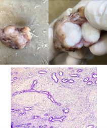 Figure 1. (A) Gross picture of excised 4x2x2 cm encapsulated nodular breast lump from right breast. (B) cut surface of right breast lump showing well-circumscribed homogeneous white fleshy area. (C) H&E-stained section at 200x showing fibroadenoma (pericanalicular pattern) with no stromal atypia.
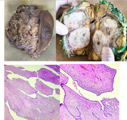 Figure 2. (A) Gross picture of left breast lump measuring 17x10x4 cm. (B) Cut section of breast lump showing well circumscribed grey white areas with cleft like spaces and cystic areas. Margins inked with green) are free grossly. (C) (D) &E stained section at 200x and 400x respectively showing stromal proliferation with mild atypia in a leaf-like pattern capped by benign ductal epithelium and intact myoepithelial layer.