Ecthyma
gangrenosum with a coinfection of methicillin-sensitive staphylococcus aureus
and streptococcus pyogenes: a case report
Rohon Das Roy 1, Dipmala Das 1,
Subhayan Das Gupta 1 *
1 Department of Microbiology, Mata
Gujri Medical College and L.S.K Hospital, Kishanganj, Bihar, India
Corresponding Authors: Subhayan Das Gupta
* Email: subspidey@gmail.com
Abstract
Introduction: Ecthyma gangrenosum
(EG) is a cutaneous infection characterized by gangrenous ulcers with
erythematous borders seen in immunocompromised as well as immunocompetent
individuals. Although Pseudomonas aeruginosa is the commonest
pathogen isolated, several other bacteria and fungi contribute to the
pathogenesis of EG. Identification of the microorganism is very essential to
initiate early empirical antimicrobial therapy.
Case presentation: We present a case report of a 13-year-old boy with multiple
recurrent ulcerative lesions in both lower extremities for the past 1 year. His
blood parameters showed signs of inflammation but was negative for aerobic
blood culture, suggesting absence of underlying bacteraemia.
There were no features of immunosuppression. On examination of pus sample,
Methicillin Sensitive Staphylococcus aureus and Streptococcus
pyogenes were isolated from the ulcerative lesions. Amoxicillin-
Clavulanate and Doxycycline was advised for 2 weeks along with surgical
debridement of the lesion followed by aseptic dressing. Patient showed complete
resolution after 2 weeks.
Discussion: Staphylococcus aureus and Streptococcus pyogenes were the causative
agents in this case, suggesting a polymicrobial association of EG besides Pseudomonas
aeruginosa. Underlying bacteraemia or any other
immunodeficiency is usually seen in a case of EG, however there are cases
reported where cutaneous manifestations show predominance.
Conclusion: A prompt diagnosis of EG is essential because there are instances when
it has proven to be fatal. Ruling out any immunodeficiency disorders and
underlying bacteraemia is of vital importance.
Administration of proper antibiotic coverage (gram positive or gram negative)
along with debridement and regular dressing can help in limiting the spread of
infections and thus improving patient outcomes.
Keywords: Ecthyma, Staphylococcus aureus, Streptococcus pyogenes
Introduction
Ecthyma gangrenosum (EG) is a cutaneous infection that
causes crusted lesions beneath which ulcers develop. It has deeper dermal
infiltration, leading to severe manifestations as compared to impetigo but both
conditions have similar bacterial causative agents. EG occurs most commonly in immunocompromised
individuals, however, healthy immunocompetent people are not always excluded.
Common risk factors include neutropenia, leukemia,
multiple myeloma, type 2 diabetes, malnutrition, and significant burn injury
(1).
Gangrenous ulcers with erythematous borders generally
characterize lesions. Primarily affecting the axillary and anogenital regions
it can also involve the arms, legs, trunk, and face. The characteristic
macroscopic appearance is caused by perivascular invasion and ischemic necrosis
of the associated skin (1).
Pseudomonas aeruginosa is the most common bacteria found in EG. P.
aeruginosa infection is rare in healthy children, but could occur in
patients with croup syndrome and sepsis. In fact, EG may be the first
sign of a Pseudomonas infection or might even develop in the
later course. It usually appears before the results of the blood culture
and help clinicians to choose appropriate antibiotics. Methicillin-resistant
Staphylococcus aureus (MRSA), Streptococcus pyogenes, Citrobacter freundii, Escherichia coli, Aeromonas hydrophila, Serratia
marcescens, Aspergillus spp., Mucor spp., and Candida
spp. are among the many other causes of EG (2).
This report suggests that besides Pseudomonas
aeruginosa, EG due to coinfection with other microorganisms, such as Staphylococcus
aureus and Streptococcus pyogenes even though rare, can prove to be
a significant finding, especially in the absence of bacteraemia or any other
immunocompromised status. Hence, prompt diagnosis with early initiation of
appropriate antibiotics can prevent further complications and fatalities.
Case presentation
We present the case of a 13-year-old boy with
complaints of multiple recurrent ulcerative lesions in both lower extremities
for the past 1 year. The lesions were itchy and slightly painful. Throughout
the past year, on application of topical ointments, there was temporary
remission of lesions which later flared up. There was no history of any insect
bite. Local examination revealed that
the lesions were in varied stages of development. Some exhibited pustules, while
others had punched-out ulcers with thick, brown-black crusts and surrounding
erythema (Figure 1). His physical examination revealed mild anaemia but no
local lymphadenopathy.
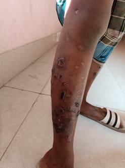
Figure 1. Multiple punched-out ulcerative lesions with thick, brown-black crusts
and surrounding erythema over lower extremities.
His blood parameters revealed mildly raised WBC count
of 13000/μL (reference value: 4000–11,000/μL), ESR 55 mm/hr (reference value: 0-15 mm/hr), CRP 150
mg/dl (reference value: ≤0.8 mg/dL), and procalcitonin 6.25 ng/ml (reference
value: ≤0.10 ng/mL). (All reference values were taken from American Board of
Internal Medicine Laboratory Test Reference Ranges ̶ July 2023). All
serological parameters were negative. Aerobic Blood culture was negative after
5 days of incubation in BD BACTECTM FX40.
Skin biopsy was taken as well as pus collected from
underneath the crusts. Gram stain of the pus revealed plenty of pus cells with
gram-positive cocci in chains as well as in clusters. Ziehl-Neelsen
staining with 20% H2SO4 was negative for acid fast
bacilli. Two cultures were done on Blood agar, one incubated aerobically at 37˚
C, and the other incubated in the presence of 10% CO2. After
overnight incubation, Staphylococcus aureus and Streptococcus
pyogenes were isolated. Antibiotic susceptibility testing was performed by
Modified Kirby Bauer Disc Diffusion method on Mueller Hilton agar for Staphylococcus
aureus and Mueller Hilton agar with 5% Sheep blood for Streptococcus
pyogenes as per CLSI 2023 guidelines. Zone diameters were measured (3). Staphylococcus
aureus was Penicillin and Clindamycin resistant, intermediate susceptible
to Ciprofloxacin and susceptible to Linezolid, Erythromycin, Cefoxitin,
Doxycycline and Cotrimoxazole. Streptococcus pyogenes was resistant to
Clindamycin but susceptible to penicillin, erythromycin, and linezolid.
Skin biopsy revealed inflammatory cell infiltration, vascular
proliferation, extensive keratinocyte necrosis along with cocci in clusters and
in chains. However, no bacilli,
amastigote forms (Leishman Donovan bodies) or fungal hyphae were found.
The patient underwent debridement of the ecthyma
crusts along with a 14 day oral course of
Amoxycillin-Clavulanate (625 mg thrice daily) and Doxycycline (100 mg twice
daily). On follow-up examination of the patient after 2 weeks, no new lesions
were seen and there was resolution of the debrided ulcers. The patient was
advised to maintain strict hygiene of the affected sites and his parents were
counselled to ensure proper nutrition of the child.
Discussion
Few differential diagnoses of EG includes other causes
of necrotic wounds such as, cutaneous anthrax, cutaneous aspergillosis,
cutaneous leishmaniasis, Mycobacterium marinum infection
and pyoderma gangrenosum (4). However, absence of bacilli, amastigote forms of
Leishmaniasis or septate hyphae fungal in the pus sample as well as skin biopsy
eliminates the first three differentials. Acid fast stain was negative for
Mycobacterial infections and absence of any relevant underlying conditions,
such as inflammatory bowel disease excludes pyoderma gangrenosum. EG is also
often confused with Ecthyma contagiosum which is characterized by
solitary pustular lesions on hands and results from the direct contact of damaged
skin with animals infected by a virus of Parapoxvirus
genus: Orf virus (5).
The diagnosis of EG is not excluded even if blood
culture yields a negative result. Pus, tissue, and exudate cultures could be
used for identifying the organism causing the lesion. When both cultures show
negative results, histopathological examination and KOH mount should be
performed.
EG is usually due to Pseudomonas aeruginosa bacteraemia
in patients with impaired immune systems. However, patients without any
underlying immunodeficiencies may also suffer from this clinical situation and
even without any features of bacteraemia (6,7,8). This is highlighted in our
case where EG occurred in an immunocompetent patient without bacteraemia and
with causative organisms besides Pseudomonas aeruginosa as Coinfection
with Methicillin Sensitive Staphylococcus aureus (MSSA) and Streptococcus
pyogenes was seen in this case. Ecthyma gangrenosum secondary to MSSA was
also seen in a case reported by Ivanaviciene J et al.
(9).
Here, the Staphylococcus
aureus strain was resistant to penicillin, whereas
the beta-haemolytic Streptococcus pyogenes was susceptible. Oral
combination antimicrobial therapy with Beta lactam-beta lactamase inhibitor
(BL-BLI) and a broad-spectrum antibiotic was required to manage this condition.
Kudo Nagata Y et al. reported cases of EG with MRSA strains, which could be
fatal, especially in patients with haematological malignancies due to
concurrent bacteraemia. Although such a case is relatively uncommon,
tissue cultures with an initial gram stain is essential for selecting
appropriate empirical antimicrobials, including the coverage of S.
aureus (10). Ulpiano Trillig, A et al. also
reported two cases of coinfection by group A Streptococcus spp. and Staphylococcus
aureus admitted to the hospital. The first patient had no risk factors nor
any immunodeficiency, but the second case was a homeless man with drug and
alcohol abuse and advanced HIV infection (11). A study in Japan showed that
staphylococcal infection was responsible for 60% of cases of EG, while the
remaining cases were attributed to Streptococcal and P. aeruginosa infections,
in descending order of prevalence (12).
There are even two postulated mechanisms identified in
the literature that describe the pathogenesis of EG. In the first form,
bacteria from a primary infection originating in the genitourinary,
respiratory, or gastrointestinal tract travel hematogenously,
disseminating through the vasculature to the skin, or in the second scenario a
cutaneous abnormality emerges and microbial infiltration takes place at the
precise location of the abnormality (13). Lesions usually recover after
surgical debridement of the ulcers with a complete course of antibiotics.
Maintenance of proper hygiene is also required to prevent recurrence.
Conclusion
Ecthyma gangrenosum is a serious and sometimes fatal
skin condition that initially manifests as a maculopapular rash, followed by a
haemorrhagic bulla, necrotic ulceration, and surrounding erythema. The
perivascular bacterial invasion of cutaneous blood vessels resulting in
ischemic skin necrosis is the main pathology behind EG. A clinical diagnosis is
often established by punched-out ulcers with thick, brown-black crusts. Lesions might be one or more, and, as seen in
our case, they can be in different phases of development. There are several
bacterial agents responsible for this condition and thus it might sometimes be
polymicrobial. Proper antibiotic therapy along with hygiene maintenance is
essential to treat this skin condition.
Ethical
consideration and
consent
Ethical
clearance was obtained from Institutional Ethical Committee. Informed written
consent was obtained from the patient to publish this case report (MGM/PRI/GEM-86/2024).
Author
contribution
RDR was responsible for conceptualization and writing the original draft. DD
contributed to the methodology, supervision and reviewing the manuscript. SDG
helped in writing and reviewing the original draft and data curation.
Conflict
of interest
The
authors declare that they have no competing interests.
Funding
There
is no funding agency involved in this research.
References
1. Vaiman M, Lazarovitch T, Heller L, Lotan G. Ecthyma gangrenosum and
ecthyma-like lesions: review article. Eur J Clin Microbiol Infect Dis. 2015
Apr; 34(4): 633-39.
2. Fang LC, Peng CC, Chi H, Lee KS, Chiu NC.
Pseudomonas aeruginosa sepsis with ecthyma gangrenosum and pseudomembranous pharyngolaryngitis in a 5-month-old boy. J Microbiol Immunol Infect. 2014 Apr; 47(2): 158-61.
3. CLSI. M100TM Performance Standards for
Antimicrobial Susceptibility Testing, 33rd ed. CLSI supplement
M100.Clinical Laboratory Standards Institute; 2023.
4. Ozkaya O, Uscetin I, Egemen O. Reconstructive procedure of lower lip
defect due to ecthyma gangrenosum: a rare complication of acute lymphoblastic leukemia. J Craniofac Surg. 2012; 23: E182–184.
5. Mavridou K, Bakola M. Orf (ecthyma
contagiosum). Pan Afr Med J. 2021; 38:322.
6.Koo SH, Lee JH, Shin H, Lee JI. Ecthyma gangrenosum
in a previously healthy infant. Arch
Plast Surg. 2012; 39: 673–75.
7. Yan W, Li W, Mu C, Wang L. Ecthyma gangrenosum and
multiple nodules: Cutaneous manifestations of Pseudomonas aeruginosa sepsis in
a previously healthy infant. Pediatr
Dermatol. 2011; 28: 204–5.
8. Goolamali SI, Fogo A,
Killian L, Shaikh H, Brathwaite N, Ford-Adams M, et al. Ecthyma gangrenosum: an important feature of pseudomonal
sepsis in a previously well child. Clin Exp Dermatol. 2009 Jul; 34(5): E180-2.
9. Ivanaviciene J, Chirch L, Grant-Kels JM, Kerr PE,
Finch J. Ecthyma gangrenosum secondary to methicillin-sensitive Staphylococcus
aureus. Int J Womens Dermatol. 2016 Jul 22; 2(3):
89-92.
10. Kudo Nagata Y, Sekiya N, Fukushima K, Horiuchi M,
Doki N. Ecthyma gangrenosum caused by Staphylococcus aureus in hematological malignancies: Case reports and literature
review. Medicine (Baltimore). 2022 Aug 19;101(33): E30070.
11. Ulpiano Trillig A, Miendje Deyi VY, Youatou P, Konopnicki D. Echtyma
gangrenosum caused by coinfection with group A Streptococcus and Staphylococcus
aureus: an emerging etiology? Case reports and
literature review. Acta Clinica Belgica. 2019; 76(1):
53–57.
12. Tomoaki I, Yoshihiko S, Maki T, Natsuko D. Ecthyma
Gangrenosum-Like Lesions in a Healthy Child after Infection Treated with
Antibiotics. Pediatr. Dermatol.2005; 22:
453-456.
13. Hajaj H, Bahari H,
Zahiri H, Ghanam A, El Ouali A, et al. Ecthyma
Gangrenosum in Patient with Bone Marrow Aplasia: A Case Report and Review of
the Literature. Open J. Pediatr. 2024;14:
272-278.