Long term clinical outcomes in patients with rectal
adenocarcinoma treated at a Tertiary Care Centre in South India
Amirthvarshan A 1,
Lijeesh A L 1, Roshni S 1*,
Mulla P A 2, Sivanandan C D 1,
Sajeed A 1, Arun Sankar 1, Geethi M H 1, Aleyamma Mathew 3
1 Department of Radiation Oncology, Regional Cancer Centre,
Thiruvananthapuram, India
2 Department of Radiation Oncology, Mangalore Institute of Oncology, Mangaluru, Karnataka, India
3 Division of Cancer epidemiology and Biostatistics, Regional cancer
Centre, Thiruvananthapuram, India
* Corresponding Author: Roshni
S
* Email: roshnisyampramod@gmail.com
Abstract
Introduction: Treatment of rectal adenocarcinoma involves multi-disciplinary approach
which includes radiation, surgery and chemotherapy. Our study aims to assess
the clinical outcomes of patients with rectal adenocarcinoma treated in a
tertiary care centre in South India.
Materials and methods: A retrospective content-based analysis was made of 131 patients
diagnosed and treated for adenocarcinoma rectum in a tertiary care hospital
during the period of 1st January 2014 to 31st December 2015. The primary
objectives were to assess disease free survival (DFS) and overall survival
(OS).
Results: Of the 131 patients, 82 were males and 49 were females with a median
age of 59 years. Stage II and Stage III disease together contributed to 65.6%
of study population. After a median follow up of 105.8 months, the 8-year
overall OS and DFS were found to be 77.2% and 78.8% respectively. In the
univariate analysis, stage of the disease and histology were found to be
significant factors in determining the overall survival (p<0.05).
Multivariate analysis showed poorly differentiated adenocarcinoma emerged as
significant factor affecting overall survival. About 31 (23.6%) patients showed
disease recurrence. Of the 31 patients, pelvic recurrence occurred in 26%
patients and among the distant metastases, liver was the most common site (33%)
followed by lung (22%).
Conclusion: The disease free survival and overall
survival of our patients were comparable with the available published data.
Keywords: Rectal cancer, Histology, Young adults, Survival rates, Prognosis
Introduction
Rectal cancer had been primarily considered a disease of the elderly,
which mostly occur after the fifth decade of life but younger people aged less
than 50 years diagnosed with colorectal cancer have been on the rise recently
(3). Most of the patients present with locally advanced disease having a 5-year
survival of 73% as per National Cancer Institute's (NCI's) Surveillance,
Epidemiology, and End Results (SEER) program and the Centers
for Disease Control and Prevention's (CDC's) National Program of Cancer
Registries (4). But the 5-year survival rate of locally advanced rectal cancer
in India is 40% which is one of the lowest in the world (5).
The standard
treatment in early-stage disease is primary surgery with or without adjuvant
treatment. In locally advanced disease, the standard of care is neoadjuvant
treatment followed by surgery with or without adjuvant chemotherapy. Assessment
of survival is the strongest parameter in oncologic outcomes. The complexity of multidisciplinary
approach and the subtle variability in clinical decision
making warrants periodical scrutiny of treatment profiles.
A study by Patil et al., showed that most of the colorectal cancers
presents at a younger age, with an aggressive histopathology, and with
involvement of both rectum and anal canal (5). There have been only few
epidemiological studies with respect to clinical outcomes of rectal cancer in
India and with its wide cultural and socio-ethnic differences, it is imperative
to assess the demographic and clinical profile that helps in strategic
planning, early diagnosis and efficient treatment modalities. Hence this study
aims to assess the clinical outcomes of patients with rectal cancer treated in
a tertiary care centre in South India.
Materials and methods
A total of 131 patients were diagnosed and
treated from June 1st 2014 to May 31st 2015 in a tertiary
care hospital in South India. This is a retrospective analytical study with
data obtained from the Medical Records in the hospital. It was based on content
analysis where the clinical and histopathological staging, treatment and follow up
details were collected from the case records using a structured proforma.
All rectal carcinoma patients with histology
of adenocarcinoma during the study period were included in the study. Histology
other than adenocarcinoma were excluded from the study. Informed consent was
obtained from all patients and the study was approved by Institutional Review
Board (No: 09/2016/04).
Statistical analysis
All patients (n = 131) were analysed and
follow up data was updated until April 2023. The primary endpoints analysed
were disease free survival (DFS) and overall survival (OS). DFS was defined as
the period from the date of registration to the date of locoregional relapse,
distant relapse or death, whichever occurred earlier. OS was defined as the
period from the date of registration to the date of death from any cause. The
statistical analysis was done using SPSS version 20. The data was analysed and
results were tabulated using descriptive statistics. Continuous variables were expressed as
mean and standard deviation, and categorical variables as counts and
percentages. Survival curves were
generated using the Kaplan‑Meier method and statistical significance was
assessed using the log‑rank test. The risk for survival was assessed
using cox regression analysis and a p-value < 0.05 was considered significant.
Results
Baseline characteristics
From June 1st 2014 to May 31st
2015, 131 patients with biopsy proven rectal adenocarcinoma were analysed in
the present study. Baseline characteristics are shown in table 1. The median
age of the population was 59 years ranging from 24 to 91 years. There were 82
males (62.59%) and 49 females (37.41%) included in the study. Mid rectal cancer
(defined as tumors residing 5-10cm from anal verge)
was the most common occurring in 51 patients (38.93%) closely followed by lower
rectal cancer (defined as tumors residing <5cm
from anal verge) diagnosed in 45 patients (34.36%) and upper rectal cancer
(defined as tumors residing 10-15cm from anal verge)
in 35 patients (26.71%). Majority had cT3 disease (38.16%) followed by cT2
(30.55%) and cT4 disease (16.03%). With respect to nodal status, 72 patients
(54.96%) had node negative disease. Stage II and Stage III disease dominated
the study population each contributing to 32.82% followed by stage I disease
(27.49%).
High risk factors like obstruction,
perforation, presence of LVE (lymphovascular
emboli)/PNI (Perineural invasion) and inadequate nodal sampling, were present
in 11 patients (8.39%). High-risk pathology like poorly differentiated
carcinoma (2.29%), mucinous adenocarcinoma (12.21%) and presence of signet ring
cells (9.92%) were observed.
Treatment characteristics
Prior to reporting at our centre, 32 patients
(24.42%) received some form of oncological treatment elsewhere. Of the
remaining 99 patients, 82 patients (82.82%) received neoadjuvant
chemoradiation. Pathological Complete Response was seen in 8.39% patients.
Pathologically node-negative disease was observed in 52.7% of patients. About
88 patients received adjuvant chemotherapy, of them 75% patients were treated
with mFOLFOX-6 (5-Fluorouracil + Folinic acid + Oxaliplatin) and 14.7% patients
were treated with CAPOX (Capecitabine + Oxaliplatin) regimen.
Table 1. Baseline Characteristics.
Characteristics |
Number (n) |
Percentage (%) |
Age |
||
<40 |
10 |
7.6% |
40-59 |
61 |
46.56% |
60-79 |
55 |
42.04% |
>80 |
5 |
3.8% |
Sex |
||
Male |
82 |
62.59% |
Female |
49 |
37.41% |
Site of disease |
||
Upper |
35 |
26.71% |
Mid |
51 |
38.93% |
Lower |
45 |
34.36% |
Tumour characteristics |
||
T1 |
20 |
15.26% |
T2 |
40 |
30.55% |
T3 |
50 |
38.16% |
T4 |
21 |
16.03% |
Nodal Status |
||
N0 |
72 |
54.96% |
N1 |
33 |
25.20% |
N2 |
26 |
19.84% |
Stage of the disease |
||
Stage I |
36 |
27.49% |
Stage II |
43 |
32.82% |
Stage III |
43 |
32.82% |
Stage IV |
9 |
6.87% |
High risk factors |
||
Obstruction |
1 |
0.76% |
Perforation |
1 |
0.76% |
LVE/PNI |
4 |
3.05% |
Inadequate Lymph Node sampling |
5 |
3.81% |
Histology |
||
Well Differentiated |
21 |
16.04% |
Moderately Differentiated |
78 |
59.54% |
Poorly differentiated |
3 |
2.29% |
Mucinous |
16 |
12.21% |
Signet ring cell |
13 |
9.92% |
Radiotherapy |
||
NACTRT |
83 |
63.35% |
Adjuvant CRT |
17 |
12.97% |
SCRT |
3 |
2.22% |
Palliative RT |
6 |
4.58% |
Abbreviations:
LVE - lymphovascular emboli; PNI - Perineural
invasion; NACTRT – Neoadjuvant chemoradiotherapy; CRT – chemoradiotherapy; SCRT
– short course radiotherapy; RT – Radiotherapy.
The median follow up was 105.8 months (16.6
to 182.6 months). The 8-year OS and DFS was shown to be 77.2% and 78.8%
respectively. Figure 1 shows the Kaplan Meier curves for 8-year DFS and OS in
our study.
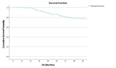
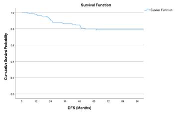
Figure 1. Kaplan Meier
curves of 8-year DFS and OS.
Tables 2 and 3 show the cumulative survival
probabilities of OS and DFS with respect to stage, sex, site of disease,
histology, and various chemotherapy regimens respectively. Figure 2 shows the
Kaplan Meier curves of OS with respect to stage, histology and nodal status.
Table 2. Survival probability with respect to OS.
Variables |
Cumulative Survival Probability (%) |
P-value |
|
Stage |
I |
77.1 |
0.009 |
II |
90.1 |
||
III |
71.9 |
||
IV |
44.4 |
||
Sex |
Female |
70.5 |
0.101 |
Male |
81.1 |
||
Age |
<50 |
72.1 |
0.371 |
>=50 |
78.9 |
||
Site |
Lower |
81.0 |
0.756 |
Mid |
76.2 |
||
Upper |
74.1 |
||
Histology |
MD |
76.2 |
<0.001 |
PD |
0.0 (all
died) |
||
WD |
84.8 |
||
Prior
Treatment |
Treated
elsewhere |
84.4 |
0.258 |
No Treatment |
74.7 |
||
Pathological
Nodal Status |
Negative |
83.6 |
0.063 |
Positive |
68.3 |
||
Adjuvant
Chemotherapy |
CAPECITABINE |
100.0 |
0.043 |
CAPOX |
61.5 |
||
FOLFOX |
81.4 |
||
Palliative
Chemotherapy |
CAPOX |
66.7 |
0.007 |
FOLFOX |
0.0 (all
died) |
||
5-FU+LV |
100.0 |
Abbreviations:
MD- Moderately Differentiated; WD- Well Differentiated; PD- Poorly
Differentiated; 5FU + LV – 5-Flourouracil + Leucovorin; mFOLFOX-6
(5-Fluorouracil + Folinic acid + Oxaliplatin); CAPOX (Capecitabine +
Oxaliplatin).
Table 3. Survival probability with respect to DFS.
Variables |
Cumulative Survival Probability (%) |
P-value |
|
Stage |
I |
80.3 |
0.775 |
II |
80.8 |
||
III |
73.7 |
||
IV |
88.9 |
||
Sex |
Female |
73.0 |
0.116 |
Male |
82.4 |
||
Age |
<50 |
75.4 |
0.434 |
>=50 |
80.0 |
||
Site |
Lower |
75.0 |
0.592 |
Mid |
77.3 |
||
Upper |
85.7 |
||
Histology |
MD |
73.8 |
0.391 |
PD |
66.7 |
||
WD |
85.7 |
||
Prior
Treatment |
Treated
elsewhere |
77.6 |
0.928 |
No Treatment |
79.2 |
||
Pathological
Nodal Status |
Negative |
75.0 |
0.473 |
Positive |
81.7 |
||
Adjuvant
Chemotherapy |
CAPECITABINE |
77.8 |
0.904 |
CAPOX |
76.2 |
||
FOLFOX |
80.2 |
||
Palliative
Chemotherapy |
CAPOX |
66.7 |
0.459 |
FOLFOX |
0.0 (all
died) |
||
5-FU+LV |
100.0 |
Abbreviations:
MD- Moderately Differentiated; WD- Well Differentiated; PD- Poorly
Differentiated; 5FU + LV – 5-Flourouracil + Leucovorin; mFOLFOX-6
(5-Fluorouracil + Folinic acid + Oxaliplatin); CAPOX (Capecitabine +
Oxaliplatin).
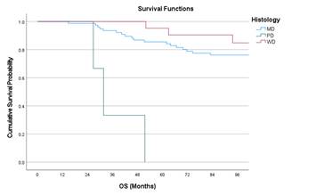
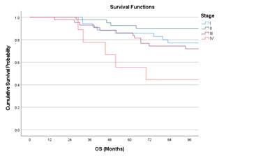
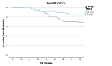
Figure 2. Kaplan Meir curves of OS with respect to Stage, Nodal status, and
histology
Univariate analysis showed stage IV had worse
outcomes compared to stage I (Hazard ratio (HR) 3.19; p = 0.042) and poorly
differentiated adenocarcinoma had worse OS compared to moderately
differentiated adenocarcinoma (HR 14.94; p = <0.001). Multivariate cox
regression analysis showed poorly differentiated histology is associated with
poor OS with an HR 9.47 (p = 0.003). Table 4 and 5 shows the univariate
analysis with respect to OS and DFS respectively. Table 6 shows the multiple
cox regression analysis affecting OS.
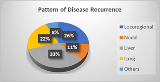
Figure 3 shows the pattern of recurrence
after radical treatment with locoregional recurrence of 26% and among the
distant sites, liver metastasis contributes to 33% followed by lung (22%),
non-regional nodes (11%) and other sites (8%) which includes bone, brain and
adrenals.
Discussion
Colorectal cancer has witnessed a staggering rise in its incidence and
parallelly the evolution of surgical and chemoradiation techniques have
improved the survival of the patients. In general, women tend to have lower
incidence and higher survival in colorectal cancer (6). Losurdo et al.,
published a gender focussed analysis of long-term outcomes of colorectal cancer
that revealed a higher 5-year (86.9% vs 80.5%) and 10-year survival (80% vs
73.3%) for women compared to men (7). This contrasts with our study where no
statistically significant survival differences were seen between men and women.
Siegel et al., reported rising incidence rates of colorectal cancer in
younger population (less than 50 years) in western countries over the last 2
decades (8). A retrospective analysis in an Indian population showed that
around 21% of patients with colorectal cancer were less than 40 years (9). Hong
et al., showed that the lowest 5-year OS and DFS were seen in age group less
than 40 years (62.5% and 52.1% respectively) (10), similar to our study where a
statistically non-significant trend of lower survival rates were seen in
patients less than 50 years.
Table 4. Univariate Cox regression analysis of OS.
Variables |
HR |
95.0% CI for HR |
Sig. |
|
Lower |
Upper |
|||
Age Group:
≥50 vs <50 |
0.70 |
0.32 |
1.54 |
0.374 |
SEX: Male vs
Female |
0.55 |
0.27 |
1.14 |
0.107 |
Stage: II vs
I |
0.41 |
0.12 |
1.36 |
0.145 |
Stage: III vs
I |
1.26 |
0.52 |
3.08 |
0.612 |
Stage: IV vs
I |
3.19 |
1.04 |
9.78 |
0.042* |
Pathological
nodal status: Yes vs No |
2.09 |
0.94 |
4.61 |
0.070 |
Adjuvant
Chemotherapy: CAPECITABINE vs FOLFOX |
0.00 |
0.00 |
0.00 |
0.981 |
Adjuvant
Chemotherapy: CAPOX vs FOLFOX |
2.68 |
0.94 |
7.60 |
0.065 |
Palliative
Chemotherapy: FOLFOX vs CAPOX |
233.29 |
0.00 |
69017143.65 |
0.396 |
Palliative
Chemotherapy: 5-FU+LV vs CAPOX |
0.05 |
0.00 |
487.19 |
0.520 |
Site: Mid vs
Lower |
1.34 |
0.55 |
3.28 |
0.522 |
Site: Upper
vs Lower |
1.39 |
0.54 |
3.60 |
0.500 |
No prior
treatment vs Treated elsewhere |
1.73 |
0.66 |
4.54 |
0.264 |
Histology: PD
vs MD |
14.94 |
4.13 |
54.06 |
<0.001* |
Histology: WD
vs MD |
0.54 |
0.16 |
1.85 |
0.329 |
Histology:
Mucin vs MD+PD+WD |
0.24 |
0.03 |
1.75 |
0.158 |
Histology:
Signet vs MD+PD+WD |
1.42 |
0.49 |
4.08 |
0.520 |
Abbreviations:
MD- Moderately Differentiated; WD- Well Differentiated; PD- Poorly
Differentiated; 5FU + LV – 5-Flourouracil + Leucovorin; mFOLFOX-6
(5-Fluorouracil + Folinic acid + Oxaliplatin); CAPOX (Capecitabine +
Oxaliplatin).
Table 5. Univariate Cox regression analysis of DFS.
Variables |
HR |
95.0% CI for HR |
Sig. |
|
Lower |
Upper |
|||
Age Group:
≥50 vs <50 |
0.72 |
0.32 |
1.65 |
0.436 |
SEX: Male vs
Female |
0.55 |
0.26 |
1.17 |
0.121 |
Stage: II vs
I |
1.00 |
0.36 |
2.75 |
0.994 |
Stage: III vs
I |
1.39 |
0.54 |
3.59 |
0.494 |
Stage: IV vs
I |
0.61 |
0.08 |
4.97 |
0.645 |
Pathological
nodal status: Yes vs No |
0.75 |
0.34 |
1.65 |
0.474 |
Adjuvant
Chemotherapy: CAPECITABINE vs FOLFOX |
1.16 |
0.26 |
5.15 |
0.844 |
Adjuvant
Chemotherapy: CAPOX vs FOLFOX |
1.32 |
0.38 |
4.62 |
0.669 |
Palliative
Chemotherapy: FOLFOX vs CAPOX |
1.41 |
0.09 |
23.57 |
0.809 |
Palliative
Chemotherapy: 5-FU+LV vs CAPOX |
0.00 |
0.00 |
0.00 |
0.981 |
Site: Mid vs
Lower |
0.88 |
0.38 |
2.03 |
0.767 |
Site: Upper
vs Lower |
0.58 |
0.20 |
1.67 |
0.314 |
No prior
treatment vs Treated elsewhere |
0.96 |
0.41 |
2.27 |
0.928 |
Histology: PD
vs MD |
1.86 |
0.25 |
13.94 |
0.548 |
Histology: WD
vs MD |
0.50 |
0.15 |
1.67 |
0.256 |
Histology:
Mucin vs MD+PD+WD |
0.51 |
0.12 |
2.16 |
0.360 |
Histology:
Signet vs MD+PD+WD |
0.32 |
0.04 |
2.37 |
0.265 |
Abbreviations:
MD- Moderately Differentiated; WD- Well Differentiated; PD- Poorly
Differentiated; 5FU + LV – 5-Flourouracil + Leucovorin; mFOLFOX-6
(5-Fluorouracil + Folinic acid + Oxaliplatin); CAPOX (Capecitabine +
Oxaliplatin).
Table 6. Multiple Cox Regression analysis for Overall OS.
Variables |
HR |
95.0% CI for HR |
Sig. |
|
Lower |
Upper |
|||
Stage: II vs
I |
0.54 |
0.16 |
1.82 |
0.316 |
Stage: III vs
I |
1.47 |
0.60 |
3.63 |
0.400 |
Stage: IV vs
I |
1.99 |
0.55 |
7.19 |
0.294 |
Histology: PD
vs MD |
9.47 |
2.10 |
42.72 |
0.003* |
Histology: WD
vs MD |
0.61 |
0.18 |
2.10 |
0.433 |
Abbreviations:
MD- Moderately Differentiated; WD- Well Differentiated; PD- Poorly
Differentiated.
Figure 3. Pattern
of disease recurrence.
Fang et al.,
reported a 5-year OS for stage I and II tumors (95.1%
and 90.3% respectively) comparable to our study where stage II had the maximal
overall survival due to lesser number of stage I patients (11). In the same
study, the survival rates dropped down to 77.8% in stage III disease and only
34.9% for stage IV disease reiterating the fact of advanced tumour stage as a
bad prognostic factor causing poor survival in patients with rectal cancer. The present study had only 9 cases with stage
IV disease constituting just 6.87% of the total population, hence a higher DFS
rate (88.9%) can be seen, but with no statistical significance.
Park et al., reported that the local relapse rates of upper rectal
cancer were 3.5% and that of mid/lower rectal cancer group were 11.1% (12). The
5-year OS of upper and mid/lower rectal cancers in the same study were 78.8%
and 73.3% respectively. This is in accordance with our study where lower rectal
cancers had a relatively poor DFS rates compared to upper rectal tumors but not statistically significant. Generally, lower rectal tumors
have a rich lymphatic drainage and vascular supply allowing for rapid and early
metastases to distant sites.
Histologically, poorly differentiated carcinoma had the worst survival
and well differentiated tumours had a better survival advantage. Most Western
studies report a prevalence of 5–15% for mucinous histology and 1% for signet
ring tumors (13,14). Vasudevan et al., reported an
inferior overall survival for signet ring cell histology compared to mucinous
adenocarcinoma (24 vs 38 months) and occurred more in younger population
compared to older adults (11% vs 4%) (15). In another study by Patil et al.,
the proportion of signet ring cell histology was higher in patients aged less
than 40 years compared to those above 40 years (22.9% vs 8.3%) (5). We observed
lower overall survival (66.7%) for patients with signet ring histology
identical to the published reports. In our multivariate analysis, poorly
differentiated histology established itself as a single most prognostic factor
affecting overall survival.
A German cohort study showed that rectal cancer patients treated in
certified cancer care centres had a better overall survival compared to those
treated in non-certified centres (5-year OS 65% vs 58.8%) owing to the high
evidence-based practices and better sophistication in certified centres (16).
The high-volume dedicated cancer centres significantly reduce in-hospital
mortality rates owing to the better quality of oncologic practices as stated in
standard guidelines. However, our study did not show a significant survival
benefit for patients treated exclusively in our centre though DFS was slightly
higher for patients treated at our centre (77.6% vs 79.2%).
Lymph node status was a single most important prognostic factor in
determining the need for adjuvant chemotherapy. Pathologically node positive
disease tends to have lower survival and frequent relapses. Diefenhardt
et al., showed that 5-year OS of 86.1% for ypN0, 74.0% for ypN1, and
43% for ypN2 and concluded that persistent lymph node metastases after
neoadjuvant therapy indicates an unfavourable phenotype of rectal cancer having
higher risk for treatment failure thus offering poor survival rates (17). This
was similar to our study where pathologically node positive population had
lower 8-year OS compared to node negative population reiterating the importance
of nodal status as a prognostic marker.
The benefit of adding oxaliplatin to standard 5-FU based chemotherapy
was first demonstrated in the MOSAIC trial as it significantly improved 5-year
DFS (73% versus 67%) and 5-year OS (79% vs 76%) (18). The phase II ADORE trial
showed addition of oxaliplatin to 5-FU after chemoRT
resulted in significantly higher 6-year DFS (68.2% vs 56.8%) and borderline
significant 6-year OS (78.1% vs 76.4%) (19). Literature showed no direct
comparisons between mFOLFOX-6 and CAPOX regime in rectal adenocarcinoma but the
comparison reports from colon cancer studies showed no significant differences
in terms of survival. Our study showed better outcomes with capecitabine and
CAPOX regimes but the sample size was too small to draw a conclusion.
The 8-year OS and DFS rates of our study were consistent with majority
of the colorectal cancer studies across the globe. Hong et al., conducted a
retrospective analysis of colorectal adenocarcinoma and reported a five-year
overall and disease-free survival of 79.5% and 69.9%, respectively (10).
Various reports from different population groups showed similar survival rates
– a Brazilian study showed 5-year OS of 60.3% (20); a Thailand single
institution study (21) showed 5-year OS and DFS of 72% and 68%
respectively and a South Australian study showed 5-year OS of 63% (22).
A noticeable observation from our study was that 25% of patients were
under 50 years of age. In the past, rectal cancer was considered as a disease
of the elderly but various Indian studies have reported a staggering rise in
early onset rectal cancers (23,24). This has been consistent across
the world where analysis of 143 million people from 20 European countries
showed CRC incidence has increased by 7.9% per year among subjects aged 20–29
years, by 4.9% among those aged 30–39 years, and by 1.6% among those aged 40–49
years from 2004 to 2016 (25). Based on
current data, it is estimated that within the next decade, 1 in 4 rectal
cancers and 1 in 10 colon cancers will be diagnosed in individuals under 50
(26). The presence of distinct genetic profiles and hereditary factors might
influence the development of early onset rectal cancers. Other factors like
inflammatory bowel disease, obesity, insulin resistance, smoking, consumption
of red meat and high fat diet, contribute to the risk of developing rectal
cancer especially in early adulthood.
The stage of the disease at diagnosis, as shown to have significant
effect on survival in our study, is crucial in determining the natural course
of the disease in terms of risk of recurrence and cancer free survival. Hence
the need for diagnosis at an early stage stands critical in the management of
rectal adenocarcinoma. The presence of tertiary care and oncology centres in
urban areas provide better health awareness to the urban population alone,
leaving the rural and low socioeconomic classes in a brim who may present at
advanced stages that significantly hamper the colorectal survival rates in our
country.
Our study has limitations. Being a retrospective study with limited
sample size, we were unable to assess toxicities of chemotherapy and
radiotherapy. Periodical evaluation of tumor markers
was not available from the records in half of the study population. We could
not analyse the survival outcomes with radiological factors like mesorectal fascia involvement (MRF), extramural vascular
invasion (EMVI) which was not routinely reported during the study period.
Surgery done from different institutions were also included in the study hence
clarity on total mesorectal excision could not be
elicited.
Conclusion
Author
contribution
Conceptualizations,
methodology, project administration, supervision, validation, review and
editing of the original draft by LA and RS; Data curation by AA,
LA, RS, MPA; Writing original draft by AA.
Investigation by LA, RS, SCD, SA, AS, GMH;
Formal analysis, software, and visualization by AM.
Ethics approval
The author
declares no conflict of interest.
Funding
There is no
funding.
References
1. Bray F, Laversanne M, Sung H, et al. Global cancer statistics 2022:
GLOBOCAN estimates of incidence and mortality worldwide for 36 cancers in 185
countries. CA Cancer J Clin. 2024; 74(3): 229-263.
2. ICMR-NCDIR.
Clinicopathological Profile of cancers in India: a report of the hospital-based
cancer registries, 2021. Accessed March 27, 2023.
3. Patra T, Mandal S, and
Alam N, et al (2018) Clinicopathological trends of colorectal carcinoma
patients in a tertiary cancer centre in Eastern India
Clin Epidemiol Glob Health 6(1) 39–43.
4. Siegel, RL, Wagle, NS, Cercek, A, Smith, RA, Jemal, A. Colorectal cancer
statistics, 2023. CA Cancer J Clin. 2023; 73(3): 233-254.
5. Patil PS, Saklani A,
and Gambhire P, et al (2017) Colorectal cancer in
India: an audit from a tertiary center in a low prevalence area Indian J Surg
Oncol 8(4) 484–490.
6. Schmuck R, Gerken M,
Teegen E, Krebs I, Klinkhammer-schalke M (2020)
Gender comparison of clinical, histopathological, therapeutic and outcome
factors in 185, 967 colon cancer patients. Langenbecks
Arch Surg. 2020 Feb;405(1):71–80.
7. Losurdo, P.,
Mastronardi, M., de Manzini, N. et al. Survival and long-term surgical outcomes
after colorectal surgery: are there any gender-related differences?.
Updates Surg 74, 1337–1343 (2022).
8. Siegel RL, Torre LA, Soerjomataram I, Hayes RB, Bray F, Weber TK, Jemal A.
Global patterns and trends in colorectal cancer incidence in young adults. Gut.
2019 Dec 1;68(12):2179-85.
9. PS A, Narayanan G, B
SS, BR KK. Colorectal Cancer in Patients Younger than 40 years -Analysis of
Clinicopathological Profile and Treatment Pattern. apjcb;9(3):301-5.
10. Hong Y, Kim J, Choi YJ,
Kang JG. Clinical study of colorectal cancer operation: Survival analysis.
Korean J Clin Oncol. 2020;16(1):3-8.
11. Fang L, Yang Z, Zhang M,
Meng M, Feng J, Chen C. Clinical characteristics and survival analysis of
colorectal cancer in China: a retrospective cohort study with 13,328 patients
from southern China. Gastroenterol Rep (Oxf).
2021;9(6):571-582.
12. Park JS, Sakai Y, Simon
NS, Law WL, Kim HR, Oh JH, Shan HC, Kwak SG, Choi GS. Long-Term Survival and
Local Relapse Following Surgery Without Radiotherapy for Locally Advanced Upper
Rectal Cancer: An International Multi-Institutional Study. Medicine (Baltimore).
2016;95(22): e2990.
13. Nitsche U, Zimmermann A,
Späth C, Müller T, Maak M, Schuster T, Slotta-Huspenina
J, Käser SA, Michalski CW, Janssen KP, Friess H,
Rosenberg R, Bader FG. Mucinous and signet-ring cell colorectal cancers differ
from classical adenocarcinomas in tumor biology and prognosis. Ann Surg. 2013
Nov;258(5):775-82.
14. Hyngstrom
JR, Hu CY, Xing Y, You YN, Feig BW, Skibber JM,
Rodriguez-Bigas MA, Cormier JN, Chang GJ. Clinicopathology and outcomes for
mucinous and signet ring colorectal adenocarcinoma: analysis from the National
Cancer Data Base. Ann Surg Oncol. 2012 Sep;19(9):2814-21.
15. Vasudevan, Smreti; Mehta, Anurag. Clinical characteristics and
survival profile of young versus old colorectal cancer patients at a tertiary
cancer center in North India over a period of 5 years. Indian Journal of Cancer
58(3): p 355-364, Jul–Sep 2021.
16. Bierbaum, V., Bobeth, C., Roessler, M. et al. Treatment in certified
cancer centers is related to better survival in patients with colon and rectal
cancer: evidence from a large German cohort study. World J Surg Onc 22, 11 (2024).
17. Diefenhardt
M, Martin D, Hofheinz R, et al. Persistent Lymph Node Metastases After
Neoadjuvant Chemoradiotherapy for Rectal Cancer. JAMA Netw
Open. 2024;7(9):e2432927.
18. André T, Boni C, Navarro
M, et al. Improved overall survival with oxaliplatin, fluorouracil, and
leucovorin as adjuvant treatment in stage II or III colon cancer in the MOSAIC
trial. J Clin Oncol 2009; 27:3109.
19. Hong YS, Kim SY, Lee JS,
Nam BH, Kim KP, Kim JE, Park YS, Park JO, Baek JY, Kim TY, Lee KW, Ahn JB, Lim
SB, Yu CS, Kim JC, Yun SH, Kim JH, Park JH, Park HC, Jung KH, Kim TW.
Oxaliplatin-Based Adjuvant Chemotherapy for Rectal Cancer After Preoperative
Chemoradiotherapy (ADORE): Long-Term Results of a Randomized Controlled Trial.
J Clin Oncol. 2019 Nov 20;37(33):3111-3123
20. Aguiar Junior S,
Oliveira MM, Silva DRM, Mello CAL, Calsavara VF,
Curado MP. Sobrevida de pacientes
com câncer colorretal em um Cancer Center. Arq
Gastroenterol. 2020;57(2):172-7.
21. Chandrachamnong,
P. (2021). Survival Analysis after Surgery of Colorectal Cancer in Vajira
Hospital. Vajira Medical Journal : Journal of Urban
Medicine, 65(4), 271–279.
22. Roder
D, Karapetis CS, Wattchow
D, Moore J, Singhal N, Joshi R, Keefe D, Fusco K, Powell K, Eckert M, Price TJ.
Colorectal cancer treatment and survival: the experience of major public
hospitals in south Australia over three decades. Asian Pac J Cancer Prev.
2015;16(6):2431-40.
23. Sudarshan V, Hussain N,
Gahine R, Mourya J. Colorectal cancer in young adults in a tertiary care
hospital in Chhattisgarh, Raipur Indian J Cancer. 2013; 50:337–40
24. Laskar RS, Talukdar FR,
Mondal R, Kannan R, Ghosh SK. High frequency of young age rectal cancer in a
tertiary care centre of southern Assam, North East
India Indian J Med Res. 2014;139:314–8
25. Vuik
F.E., Nieuwenburg S.A., Bardou M., Lansdorp-Vogelaar I., Dinis-Ribeiro M., Bento M.J., Zadnik
V., Pellisé M., Esteban L., Kaminski M., et al.
Increasing incidence of colorectal cancer in young adults in Europe over the
last 25 years. Gut. 2019;68:1820–1826.