Unusual extrahepatic sites of hepatocellular metastases: case series
Keywords:
Hepatocellular carcinoma, Extrahepatic metastases, HepPar-1Abstract
Introduction: Hepatocellular carcinoma (HCC) is a major global health burden and a leading cause of cancer-related mortality. Patients typically present with symptoms related to the primary hepatic tumor, while extrahepatic metastases are generally associated with advanced disease and poor prognosis. Rarely, HCC may initially manifest through symptoms arising from metastatic involvement before the primary hepatic lesion is identified.
Case Presentation: We report two cases of hepatocellular carcinoma that initially presented with symptoms attributable to extrahepatic metastases to the rare sites. Both cases had normal AFP levels and were CT negative, with HCC detected only on MRI of the abdomen.
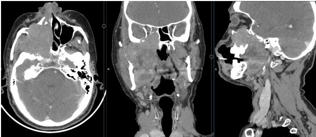
Case 1: A 63-year-old male presenting with epistaxis and a destructive maxillary sinus mass, which on histopathology and immunohistochemistry was diagnosed as metastatic HCC. Subsequent MRI of the liver revealed a small LI-RADS 5 lesion that was occult on CT.
Case 2: A 60-year-old chronic alcoholic male presenting with gastrointestinal bleeding due to a duodenal mass. Biopsy confirmed metastatic HCC, and subsequent MRI demonstrated two hepatic lesions categorized as LI-RADS 5 and LI-RADS 4, which were not definitively detected on initial CT imaging.
Discussion: Extrahepatic metastases from HCC most commonly involve the lungs, lymph nodes, and bones, while involvement of unusual sites such as the maxillary sinus and duodenum is exceedingly rare. Presentation with metastatic disease prior to identification of the primary tumor may delay diagnosis and management. These cases highlight the limitations of CT in detecting small or infiltrative hepatic lesions and emphasize the superior sensitivity of MRI in lesion characterization. Immunohistochemical markers such as HepPar-1 and arginase play a crucial role in establishing the diagnosis of metastatic HCC, particularly when serum alpha-fetoprotein levels are normal or only mildly elevated.
Conclusion: Hepatocellular carcinoma may rarely present with symptoms related to extrahepatic metastases before the primary hepatic lesion becomes clinically or radiologically apparent. A high index of suspicion, combined with comprehensive imaging using MRI and appropriate immunohistochemical analysis, is essential for early diagnosis. Recognition of such atypical presentations can prevent diagnostic delay and facilitate timely initiation of appropriate therapy.
Additional Files
Published
How to Cite
License
Copyright (c) 2025 Neha Chhabra, Puneet Somal, Sankalp Sancheti

This work is licensed under a Creative Commons Attribution-NonCommercial 4.0 International License.
