An examination of the cytomorphological trends in tuberculous lymphadenitis at a tertiary care hospital of southern Assam
Keywords:
Cytomorphological patterns, Tuberculous lymphadenitis, Ziehl-Neelsen (ZN) staining, Acid-fast bacilliAbstract
Introduction: Tuberculous lymphadenitis is a leading cause of lymph node enlargement accounting for 195 per 1,00,000 population in India. Fine needle aspiration cytology (FNAC) is rapid and economical compared to other tests and thus plays a crucial role in diagnosing this condition. It prevents unnecessary biopsy of lymph nodes and it can be used for collection of material for cytomorphological and bacteriological examination. This study aimed to assess the cytomorphological patterns of tuberculous lymphadenitis and correlate them with Ziehl-Neelsen (ZN) staining.
Primary aim is to assess the cytomorphological patterns of tubercular lymphadenitis and to correlate with the bacteriological results using ZN staining.
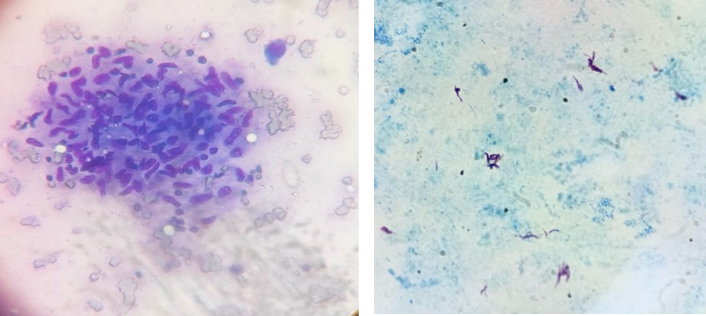
Materials and methods: The FNAC results of 100 cases diagnosed with tuberculous lymphadenitis over a period of one year were analyzed from the cytopathology section of Silchar Medical College and Hospital. The findings were classified into three patterns: pattern A - epithelioid granuloma in absence of caseous necrosis, pattern B - epithelioid granuloma with caseous necrosis, and pattern C - caseous necrosis in absence of epithelioid granuloma. The cytomorphological patterns were then correlated with acid-fast bacilli (AFB) positivity.
Results: In individuals between the ages of 21 and 30, tuberculous lymphadenitis was predominantly observed. The cervical lymph node (92%) was the most frequently affected area. Among the different patterns of the condition, Pattern B, which is characterized by the presence of epithelioid granuloma along with caseous necrosis, was found to be the most common (53%). In contrast, Pattern C, which is marked by caseous necrosis without the presence of epithelioid granuloma, exhibited the highest positivity for acid-fast bacilli (80%). The difference in AFB positivity among the patterns was statistically significant (P-value= 0.0003).
Conclusion: FNAC is an effective and economical diagnostic tool for tuberculous lymphadenitis, particularly in resource-limited settings. The study found that Pattern B (epithelioid granuloma with caseous necrosis) was the most common, while Pattern C (caseous necrosis without epithelioid granuloma) exhibited the highest AFB positivity. FNAC, combined with ZN staining, enhances the accuracy of tuberculosis diagnosis, minimizing the need for invasive biopsies. Given the high prevalence of tuberculosis, FNAC should be the first-line investigation for patients presenting with superficial lymphadenopathy, ensuring timely diagnosis and treatment.
Additional Files
Published
How to Cite
License
Copyright (c) 2025 Sukanya Choudhury, Payel Hazari, Nicky Choudhury, Shah Alam Sheikh

This work is licensed under a Creative Commons Attribution-NonCommercial 4.0 International License.
