Primary pleural B-cell lymphoma mimicking tuberculosis complicated by pulmonary embolism and suspected rituximab-induced interstitial lung disease
Keywords:
Primary pleural lymphoma, Non-Hodgkin lymphoma, Pleural effusion, Tuberculosis, Rituximab-induced lung injury, Pulmonary embolismAbstract
Introduction: Primary pleural non-Hodgkin lymphoma (PPL) is an extremely rare malignancy (<0.3% of non-Hodgkin lymphomas) that frequently masquerades as tuberculosis or mesothelioma, causing diagnostic delay in endemic regions.
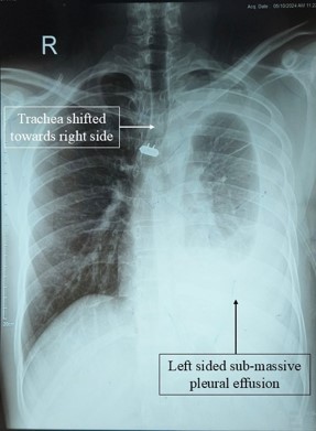
Case presentation: A 27-year-old male presented with sub-massive hemorrhagic pleural effusion and sub-centimetric pleural nodules. Pleural fluid analysis showed lymphocyte-predominant exudate with low adenosine deaminase (18 IU/L) and negative tuberculosis tests. Contrast-enhanced computed tomography and thoracic ultrasound demonstrated nodular pleural thickening. Cytology was discordant between cell block (suggesting lymphoma) and fine-needle aspiration (suggesting small-cell lung carcinoma). Immunohistochemistry resolved the diagnosis, showing positivity for CD45 and CD20, confirming B-cell non-Hodgkin lymphoma. Flurodeoxyglucose (FDG) Positron Emission Tomography demonstrated disease apparently confined to the pleura without nodal or distant organ involvement. The patient received R-CHOP chemo-immunotherapy. Two months later, he developed severe hypoxemia. Computed tomography pulmonary angiography revealed extensive GGOs and interlobar septal thickening consistent with rituximab-induced interstitial lung disease alongside left main pulmonary thromboembolism. Despite high-dose methylprednisolone and anticoagulation, the patient succumbed to respiratory failure.
Discussion: PPL should be considered in patients presenting with hemorrhagic pleural effusion, low pleural fluid adenosine deaminase levels, and pleural nodularity, even in young individuals. When cytological findings are inconclusive or conflicting, pleural tissue sampling and immunohistochemistry are essential for establishing the diagnosis. This case also highlights the potential for life-threatening treatment-associated complications, including suspected rituximab-induced pneumonitis and concurrent pulmonary thromboembolism.
Conclusion: Primary pleural B-cell non-Hodgkin lymphoma is a diagnostic challenge because of its rarity and nonspecific clinical presentation. Early tissue diagnosis with immunohistochemical confirmation is crucial. Clinicians should also maintain a high index of suspicion for severe pulmonary complications during treatment, as concurrent interstitial lung disease and pulmonary thromboembolism may have fatal consequences.
Additional Files
Published
How to Cite
License
Copyright (c) 2026 Saikat Bhowmik, Thaodem Collin Singh, Sunanda Haorongbam, Tarunkumari Thoudam

This work is licensed under a Creative Commons Attribution-NonCommercial 4.0 International License.